Google+Linking To And Excerpting From "2022 ACC Expert Consensus Decision Pathway on the Evaluation and Disposition of Acute Chest Pain in the Emergency Department" - Tom Wade MD
Linking To And Excerpting From “2022 ACC Expert Consensus Decision Pathway on the Evaluation and Disposition of Acute Chest Pain in the Emergency Department”
Today, I review, link to, and excerpt from 2022 ACC Expert Consensus Decision Pathway on the Evaluation and Disposition of Acute Chest Pain in the Emergency Department [No abstract available] [Full-Text HTML] [Full-Text PDF]. J Am Coll Cardiol. 2022 Oct 11;80(20):1925–1960. doi: 10.1016/j.jacc.2022.08.750
The objectives of this Expert Consensus Decision Pathway are to provide structure around the evaluation of chest pain in the ED and to facilitate rapid disposition and limit unnecessary testing among patients with chest pain who are at low risk and who do not have ACS. The document also aims to provide critical appraisal of the options for clinical decision pathways (CDPs) that hospitals may choose from to achieve these aims. Implementation of accelerated CDPs has the potential to further reduce ED length of stay and increase the proportion of patients who are eligible for rapid ED discharge and do not routinely require additional diagnostic testing, without compromising patient safety.
To limit inconsistencies in interpretation and to develop guidance that is complementary to current evidence-based guidelines for the management of chest pain in the ED, specific definitions and assumptions were considered by the writing committee in the development of the consensus recommendations.
3.1. Definitions
See article.
3.2. General Clinical Assumptions
The content of this Expert Consensus Decision Pathway applies only to patients presenting to the ED with chest pain or other symptoms suggestive of myocardial ischemia undergoing evaluation for possible ACS. The Expert Consensus Decision Pathway does not apply to patients with stable angina or those evaluated in settings other than the ED. For these other patient groups, the 2021 AHA/ACC/multisociety chest pain guideline provides comprehensive recommendations on chest pain evaluation and management not limited to the ED setting.5 This Expert Consensus Decision Pathway is not applicable to patients with hemodynamic instability, significant heart failure, or other conditions that would mandate hospital admission.
The document is focused on the rapid evaluation and disposition of patients with possible ACS in the ED. It does not address the evaluation and management of patients with definite ACS or to serve as a guide for the diagnosis or management of MI. Readers are referred to the 2013 ACCF/AHA Guideline for the Management of ST-Elevation Myocardial Infarction (STEMI),12 2014 AHA/ACC Guideline for the Management of Patients with Non-ST-Elevation Acute Coronary Syndromes (NSTE-ACS),13 and the Fourth Universal Definition of MI14 for comprehensive recommendations on these topics.
This Expert Consensus Decision Pathway is focused on CDPs using high-sensitivity cardiac troponin I (hs-cTnI) assays. The pathways are not appropriate for use with older-generation, less-sensitive assays. An important secondary objective of this document is to support the transition to hs-cTn assays, which offer important advantages for the rapid evaluation and disposition of chest pain in the ED and are recommended by the 2021 AHA/ACC/multisociety chest pain guideline.5 It is recommended that U.S. centers transition to hs-cTn assays for optimal patient care.
Recommendations regarding noninvasive testing should be considered in the context of availability of institutional testing and local expertise. However, coronary computed tomography angiography (CTA) is an important tool for evaluation of intermediate-risk patients in the ED, and thus, broader application across centers and greater availability within centers is recommended.
CDPs for rapid evaluation of chest pain should be interpreted within the context of all available clinical information. The provider’s clinical judgment at the bedside remains an indispensable tool that may lead to different triage decisions than those suggested by the CDP.
The rule-out cutpoints for high-sensitivity cardiac troponin T (hs-cTnT) and hs-cTnI assays recommended in this document may differ slightly from those reported in studies evaluating accelerated CDPs, because we have synthesized data across multiple studies and used concentration cutoffs permitted for reporting in the United States by the FDA. The CDPs included in this document use the LoQ (see definition in the previous text) permitted by the FDA for the 0-hour rule-out criterion.
The recommendations in this document are based on available data, much of which is observational rather than from randomized controlled trials. As new, relevant, and sound data become available, modifications to CDPs may be necessary.
Successful implementation of the CDPs outlined in this document requires engagement of a multidisciplinary team with collaboration among emergency medicine, laboratory medicine, cardiology, and hospital medicine specialties. Clinicians caring for patients in whom hs-cTn assays are used need to be aware of the clinical decision thresholds as well as the strengths and limitations of the CDP. Specific recommendations for transitioning to hs-cTn assays have been outlined previously.15
4. SUMMARY GRAPHIC
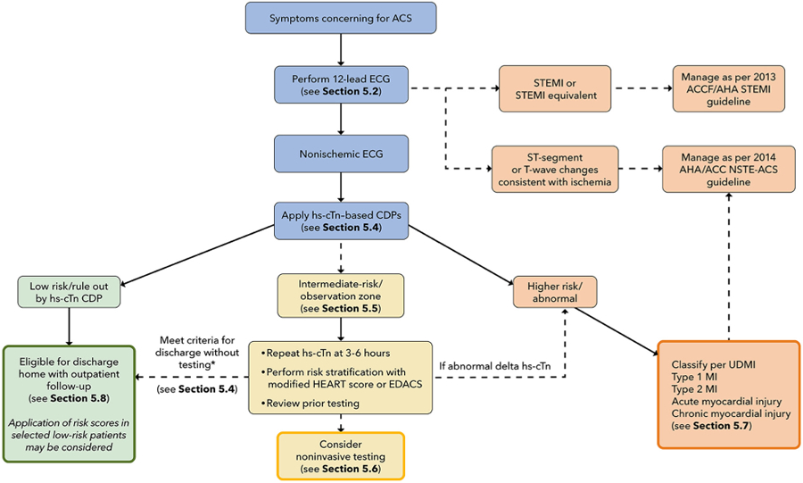
This Expert Consensus Decision Pathway is designed to parallel the usual course of evaluation of patients in the ED with symptoms requiring evaluation for possible ACS (see Figure 1). The first step is careful evaluation of the ECG (see Section 5.2). Patients with a nonischemic ECG can enter an accelerated CDP designed to provide rapid risk assessment and exclusion of ACS (see Section 5.4). Patients classified as low risk (rule out) using hs-cTn–based CDPs supported by this document can generally be discharged directly from the ED without additional testing, although outpatient testing may be considered in selected cases. In contrast, patients with substantially elevated initial hs-cTn values or those who have significant dynamic changes over 1 to 3 hours are assigned to the abnormal/high-risk category and should be further classified according to the Universal Definition of MI into type 1 or 2 MI or acute or chronic nonischemic cardiac injury (see Section 5.7). High-risk patients should usually be admitted to an inpatient setting for further evaluation and treatment. Patients determined to be intermediate risk with the CDP should undergo additional observation with repeat hs-cTn measurements at 3 to 6 hours and risk assessment using either the modified History, ECG, Age, Risk Factors, and Troponin (HEART) score or the ED Assessment of Chest Pain Score (EDACS) (see Section 5.5). Noninvasive testing should be considered for the intermediate-risk group unless low-risk features are identified using risk scores or noninvasive testing has been performed recently with normal or low-risk findings. Details of the assessment steps are provided in Section 5.
*Unchanged high-sensitivity troponin concentration (ie, no or minimal change over serial measurements) with 1) recent normal testing (ie, invasive or CT coronary angiogram <2 years ago or stress test <1 year ago); 2) symptoms inconsistent with possible ACS; 3) chronic elevations in hs-cTn that are unchanged compared with levels measured previously; or 4) a modified HEART score ≤3 or EDACS <16.
ACC = American College Cardiology; ACS = acute coronary syndrome; AHA = American Heart Association; CDP = clinical decision pathway; ECG = electrocardiogram; EDACS = Emergency Department Assessment of Chest Pain Score; HEART = History, ECG, Age, Risk Factors, and Troponin; hs-cTn = high-sensitivity cardiac troponin; MI = myocardial infarction; NSTE-ACS = non-ST-segment elevation acute coronary syndrome; STEMI = ST-segment elevation myocardial infarction; UDMI = Universal Definition of MI.
The initial clinical evaluation of a patient with acute chest pain should focus on rapid identification and treatment of patients with life-threatening conditions such as ACS, aortic dissection, and pulmonary embolism. Patients who are hemodynamically unstable, have significant arrhythmias, or have evidence of significant heart failure should be evaluated and treated appropriately and are not candidates for an accelerated CDP.
Although the term chest discomfort is more accurate, the term chest pain is embedded in clinical use and will be used throughout this document to describe potential ischemic chest symptoms. Symptoms described as a pressure, tightness, squeezing, heaviness, or burning should be considered consistent with ACS. Pain locations other than the chest can also occur and include the shoulder, arm, neck, back, upper abdomen, or jaw. Other associated symptoms include shortness of breath, nausea, vomiting, diaphoresis, fatigue, and mental status changes, which, in some cases, may be the predominant symptom. In contrast, symptoms described as sharp, fleeting, related to inspiration (pleuritic) or position, or localized to a single point are unlikely to represent myocardial ischemia.
Chest pain has been traditionally classified as “typical” or “atypical.” The 2021 AHA/ACC/multisociety chest pain guideline discourages the use of the term atypical chest pain and instead emphasizes focusing on specific aspects that suggest whether the pain is likely related to ischemia. The guideline also recommends using cardiac, possible cardiac, and noncardiac to describe the suspected cause of chest pain.5 Chest pain should be considered stable when symptoms are chronic and associated with precipitants such as exertion or emotional stress.
Clinical assessment should include the chest pain description and associated symptoms, onset, duration, location, radiation, and precipitating and relieving factors. In addition, a detailed assessment of cardiovascular risk factors and medical, family, and social history should complement the assessment of presenting symptoms. The results of prior testing for coronary artery disease (CAD) as well as prior computed tomography (CT) imaging of the chest that delineates the presence and severity of coronary calcification should be reviewed, as it may inform subsequent diagnostic testing strategies.
There are no physical examination findings specific for coronary ischemia; thus, the examination should be targeted to identify findings associated with high risk for morbidity and mortality in ACS, or to the presence of potential alternative diagnoses. High-risk examination findings include signs of low cardiac output (tachycardia, hypotension, cool extremities, low urine output, and altered mental status), heart failure (pulmonary edema, elevated jugular venous pressure, and peripheral edema), and a new systolic murmur concerning for acute mitral regurgitation or a ventricular septal defect. Clues to non-ACS causes for the patient’s symptoms include fever (endocarditis, pneumonia), pulse differential (aortic dissection), abnormal lung findings (pneumonia, pneumothorax), and abnormal cardiac findings such as a pericardial friction rub (pericarditis) or other murmurs (aortic stenosis, outflow tract obstruction, endocarditis).
As part of the initial assessment, a chest X-ray should be performed in almost all patients with possible ACS, given the potential to identify high-risk findings such as pulmonary edema, as well as to identify potential noncardiac causes for the patient’s symptoms. Performance of a chest X-ray should not delay emergent interventions such as primary percutaneous coronary intervention for those with a definitive STEMI.
5.2. Initial Evaluation: Focus on ECG
5.2.1. Initial ECG Interpretation
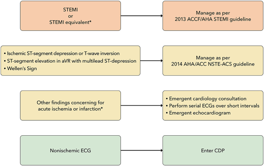
The ECG is critical for the initial assessment and management of patients with potential ACS and therefore should be performed and interpreted within 10 minutes of arrival at the ED.5,12,13 In patients who arrive via emergency medical service transport, the pre-hospital ECG should be reviewed, because ischemic changes may have resolved before ED arrival. In the ED, the initial ECG should be examined for signs of ischemia (see Figure 2), particularly for STEMI or a STEMI equivalent (see Table 1), as this identifies patients who should undergo immediate reperfusion therapy and be managed in accordance with the 2013 STEMI guideline.12 Automated ECG algorithms provide an immediate interpretation and diagnostic assistance, particularly for the inexperienced ECG reader, and may identify subtle ECG changes that just meet STEMI criteria, particularly with inferior ST-segment elevation. However, interpretation accuracy varies among different algorithms, with up to a 2-fold variation in identification of ECGs concerning for ACS.16 Unfortunately, physician accuracy for determining ischemic ECG changes is also variable, with lower sensitivity for smaller degrees of ST-segment elevation.17
*See Table 1 for ECG findings of STEMI equivalent and findings consistent with ischemia or infarction.
ACC = American College of Cardiology; AHA = American Heart Association; aVR = augmented vector right; CDP = clinical decision pathway; ECG = electrocardiogram; NSTE-ACS = non–ST-segment elevation acute coronary syndrome; STEMI = ST-segment elevation myocardial infarction.
TABLE 1.
Electrocardiogram Findings Suggestive of Ischemia
FINDING
CRITERIA
STEMI equivalents
Posterior STEMI
Criteria:
Horizontal ST-segment depression in V1-V3
Dominant R-wave (R/S ratio >1) in V2
Upright T waves in anterior leads
Prominent and broad R-wave (>30 ms)
Confirmed by:
ST-segment elevation of ≤0.5 mm in at least 1 of leads V7-V9*
Left bundle branch block or ventricular paced rhythm with Sgarbossa Criteria
A total score ≥3 points is required:
Concordant ST-segment elevation ≥1 mm in leads with a positive QRS complex (5 points)
Concordant ST-segment depression ≥1 mm in leads V1-V3 (3 points)
Discordant ST-segment elevation ≥5 mm in leads with a negative QRS complex (2 points)
If there is discordant ST-segment elevation ≥5 mm, consider ST/S ratio <−0.25
Left bundle branch block or ventricular paced rhythm with Smith-modified Sgarbossa Criteria
Positive if any of the following are present:
Concordant ST-segment elevation of 1 mm in leads with a positive QRS complex
Concordant ST-segment depression of 1 mm in V1-V3
ST-segment elevation at the J-point, relative to the QRS onset, is at least 1 mm and has an amplitude of at least 25% of the preceding S-wave
De Winter Sign
Tall, prominent, symmetrical T waves arising from upsloping ST-segment depression >1 mm at the J-point in the precordial leads
0.5–1 mm ST-segment elevation may be seen in lead aVR
Hyperacute T waves
Broad, asymmetric, peaked T waves may be seen early in STEMI
Serial ECGs over very short intervals are useful to assess for progression to STEMI
ECG findings consistent with acute/subacute myocardial ischemia
aVR ST-segment elevation
Most often caused by diffuse subendocardial ischemia and usually occurs in the setting of significant left main coronary artery or multivessel coronary artery disease
ST-segment elevation in aVR ≤1 mm
Multilead ST-segment depression in leads I, II, Val, and/or V4-V6
Absence of contiguous ST-segment elevation in other leads
ST-segment depression
Horizontal or downsloping ST-segment depression ≥0.5 mm at the J-point in 2 or more contiguous leads is suggestive of myocardial ischemia
Wellen’s syndrome
Clinical syndrome characterized by:
Biphasic or deeply inverted and symmetric T waves in leads V2 and V3 (may extend to V6)
Recent angina
Absence of Q waves
Inverted T waves
May be seen in ischemia (subacute) or infarction (may be fixed and associated with Q waves) in continuous leads
V7 placed at left posterior axillary line in same plane as V6; V8 placed at the tip of the left scapula; V9 placed in the left paraspinal region in the same plane as V6. aVR = augmented vector right; ECG = electrocardiogram; STEMI = ST-elevation myocardial infarction.
In the absence of ischemic ST-segment elevation, the ECG should be examined for other changes that have been associated with coronary artery occlusion (see Table 1)18–21; when present, these should prompt evaluation for emergent coronary angiography.
For patients suspected of ACS who have ST-segment or T-wave changes suggestive of ischemia, comparison with previous ECGs can be helpful.22 Emergent consultation for expert over-read should be obtained for ECGs concerning for ACS that lack clear diagnostic criteria. Serial ECGs performed over short time intervals in those with a high suspicion for ACS may detect dynamic ischemic changes.23,24 ECGs performed later, even the next day, may show evolution of findings that confirm the diagnosis, such as Q waves or new T-wave inversions. A posterior ECG should be performed if the initial ECG is nondiagnostic but suspicion for a posterior MI is high (see Section 5.2.2 and Table 1). Emergent transthoracic echocardiography (TTE) for assessment of wall motion should be considered in patients with ECGs concerning for but not diagnostic of ischemia and infarction, particularly when borderline ST-segment elevation or left bundle branch block (LBBB) or equivocal signs of posterior MI are present. Because accurate assessment of wall motion is difficult, the TTE should be performed and reviewed by a clinician qualified in echocardiography (see Section 5.6).
Finally, the ECG should be reviewed for other findings suggesting alternative causes for the patient’s symptoms, such as pericarditis or pulmonary embolism. The absence of ischemic ECG changes identifies patients at relatively lower (although not necessarily low) risk for MI and ischemic complications but is not sufficient for excluding ACS25,26; therefore, these patients are appropriate for evaluating using a CDP.
All CDPs (see Section 5.4) exclude patients with ischemic ST-segment elevation; however, many do not specifically exclude those with other ECG findings potentially associated with ischemia.27 We recommend that patients with new ischemic ECG changes should be considered high risk and undergo evaluation and treatment according to current NSTE-ACS guidelines13 and not be entered into an accelerated CDP. For the purposes of this document, ECGs are classified into 3 groups: 1) STEMI or equivalent; 2) ischemic ST-segment or T-wave abnormalities; and 3) nonischemic, which includes ECGs interpreted as normal, having nonspecific findings, left ventricular hypertrophy with or without repolarization ST-T wave changes, and left or right bundle branch block or paced rhythm (not meeting Sgarbossa9 or modified Sgarbossa10,11 criteria for MI).
5.2.2. Additional ECG Findings Consistent With Acute Coronary Artery Occlusion
The application of STEMI ECG criteria on a standard 12-lead ECG alone will miss a significant minority of patients who have acute coronary occlusion.21 Therefore, the ECG should be closely examined for subtle changes that may represent initial ECG signs of vessel occlusion, such as hyperacute T waves (in the absence of electrolyte imbalances or significant left ventricular hypertrophy) or ST-segment elevation <1 mm, particularly when combined with reciprocal ST-segment depression, as this may represent abnormal coronary blood flow and/or vessel occlusion.21 Concomitant reciprocal ST-segment depression may be visually more evident than the minor ST-segment elevation in such patients.28 If present, these patients should be evaluated for emergent coronary angiography as their outcomes are similar to those with more extensive ST-segment elevation.29
Other ECG findings may also indicate acute coronary artery occlusion. Anterior ST-segment depression (eg, leads V1-V3) may represent acute posterior transmural MI.19 Acute posterior MI can be confirmed by evaluating for the presence of ST-segment elevation on posterior leads19 or emergent echocardiography demonstrating wall motion abnormalities in the posterior and/or inferior territories. Emergent coronary angiography should be performed when there is a high suspicion for acute posterior MI, because delay in reperfusion has been associated with worse outcomes.30 Similarly, de Winter’s sign, suggested by tall, prominent, symmetrical T waves arising from upsloping ST-segment depressions >1 mm in the precordial leads, can be seen in proximal left anterior descending artery occlusion and therefore warrants immediate angiography.18
The identification of acute coronary occlusion among patients with LBBB or ventricular pacing poses particular challenges. The presence of a new LBBB is no longer considered a STEMI equivalent,12 although it is associated with higher risk because most patients with LBBB have underlying cardiac disease, typically CAD or a cardiomyopathy.31 The Sgarbossa criteria (see Table 1) are specific, although not sensitive for acute coronary artery occlusion.9 In a study that included patients with acute left anterior descending artery occlusion, a modification that used an ST/T-wave ratio improved sensitivity from 52% to 91% with similar specificity to the original criteria.10,11 For those meeting the Sgarbossa or modified Sgarbossa criteria, treatment should be similar to those with STEMI. Emergent echocardiography can be performed in cases in which there is an LBBB and suspicion for ischemia/infarction with the caveat that the frequent coexistent cardiomyopathy may make differentiation difficult. Although less well studied, patients with ventricular paced rhythms who have ECG findings that meet Sgarbossa criteria should also be considered high risk and undergo emergent coronary angiography.9,32
5.2.3. ECG Findings Consistent With Ischemia
Wellen’s syndrome is characterized by biphasic or inverted T-wave inversions in the anterior precordial leads in patients whose ischemia has resolved.33 Its presence is associated with proximal left anterior descending artery stenosis and is associated with high rate of subsequent transmural MI.
In patients with ischemic symptoms, ST-segment elevation in lead augmented vector right (with or without elevation in V1) combined with multilead ST-segment depression represents a high-risk ECG finding that is associated with high morbidity and mortality.34 In patients with ischemic symptoms, this often represents diffuse ischemia due to significant stenosis involving the left main and/or 3-vessel disease,35,36 although it can be seen in other non-ACS conditions causing a demand/supply mismatch.37 In approximately 10% of cases, acute coronary occlusion is present.35 Accordingly, management of patients with this ECG pattern must be nuanced. Precipitants of supply/demand mismatch (if present) should be treated. Emergent coronary angiography should be considered in patients with persistent ischemic symptoms or ECG changes after treatment or if there is hemodynamic instability.35
Ischemic ST-segment depression is present in a minority of ACS patients, representing a specific, but not sensitive finding for ACS. In the setting of ischemic symptoms, it should prompt treatment consistent with the 2014 NSTE-ACS guidelines,13 as the majority of patients with ischemic ST-segment depression will be diagnosed with MI.38 T-wave inversion is a less-specific marker of subendocardial ischemia because it can be present in non-ACS conditions. Rather than being indicative of acute ischemia, T-wave inversion can become evident after clinical ischemia resolves. Ischemic T-wave inversion tends to be deeper and new when compared with prior ECGs.
5.2.4. Summary
The ECG is a critical component of the initial assessment and management of ED patients with possible ACS. The ECG should be rapidly assessed for evidence of acute infarction or ischemia, and if present, subsequent care should follow current guidelines for management of acute STEMI12 and NSTE-ACS.13 The ECG should also be examined for subtle changes that are also consistent with ACS as well as for other findings that could suggest a non-ACS cause for their symptoms. Patients who have a nonischemic ECG (as defined previously) are eligible for entering a CDP, and further clinical evaluation should take place as outlined later.
5.3. Hs-cTn Assays
Measurement of cardiac troponin T (cTnT) or cardiac troponin I (cTnI), the gold-standard biomarkers of myocardial injury, is critical for the evaluation of possible ACS in the ED. In the United States, hs-cTn assays are being increasingly adopted because of their ability to detect lower cTn concentrations with improved analytical performance (ie, greater sensitivity and precision) compared with older-generation assays.15 “High sensitivity” in this context refers to assay characteristics; the analytes being measured (cTnT and cTnI) are the same as for older-generation, conventional assays. Accordingly, hs-cTn assays are preferred to conventional assays for the evaluation of acute chest pain.5,6 The “high-sensitivity” designation was initially adopted by cTn assay manufacturers in the absence of clear analytical criteria defining high sensitivity. The International Federation of Clinical Chemistry and Laboratory Medicine Task Force on Clinical Applications of Cardiac Biomarkers lists 2 criteria to define “true” hs-cTn assays: first, the assay imprecision (ie, CV) at the 99th percentile value for that assay should be ≤10%; and second, at least 50% of apparently healthy men and women7 should have cTn concentrations above the assay’s LoD.39 Most manufacturers now voluntarily report assay characteristics according to these criteria (see Table A in the Supplemental Appendix). Of note, point-of-care cTn assays must also meet these criteria to be considered high-sensitivity assays.
When considering assay characteristics, it is important to understand the terminology commonly used both in product inserts and in the literature. The LoB is the highest apparent cTn concentration found with a given assay when testing replicates of a sample known to contain no cTn (ie, blank sample). The LoD is the lowest cTn concentration that can be reliably distinguished from the LoB when testing replicates of samples known to contain cTn. In research settings, the term undetectable troponin is typically used for cTn concentrations that are below the LoD of the assay. The LoQ is the lowest cTn concentration that can be reported reliably as a number, either with a CV ≤20% (as per FDA regulations), or with a more stringent CV ≤10%. Analytical definitions are summarized in Figure A of the Supplemental Appendix.15 In clinical settings, the FDA only allows cTn concentrations to be reported numerically if they are equal to or above the LoQ; therefore, cTn concentrations above the LoD but below the LoQ are not reported in U.S. clinical settings. This has implications for algorithms that use a single hs-cTn measurement at ED arrival to exclude MI. For example, for hs-cTnT, the lowest value that the FDA allows to be reported is 6 ng/L, although the LoD is 3 to 5 ng/L depending on the specific analyzer used. It is therefore important to interpret the term undetectable troponin in context for each hs-cTn assay and for each usage scenario.
Concentrations of hs-cTn should be reported in whole numbers without decimal places as nanograms per liter (ng/L), as recommended by the International Federation of Clinical Chemistry and Laboratory Medicine, endorsed in the Fourth Universal Definition of MI,14,39 and adhered to by assay manufacturers. Reporting in ng/L avoids potential confusion related to the use of 3 decimal places and multiple zeroes (for instance, 14 ng/L is preferred over 0.014 ng/mL).
5.3.1. Defining Abnormal Hs-cTn Values
Key questions for the management of patients with possible ACS in the ED are what constitutes an “abnormal” or an “elevated” hs-cTn value and how to rapidly and reliably differentiate between ACS and the multitude of other potential causes for cTn elevation (see Section 5.7).40 In the absence of an objective cTn threshold, the 99th percentile URL cTn value derived from a “normal reference population” has been endorsed by expert consensus as a cutpoint for the diagnosis of MI for more than 20 years.41 However, studies in large population-based cohorts have shown that hs-cTn represents a continuum of risk, such that minor cTn elevations (detectable but below the 99th percentile URL) are associated with structural heart disease, worse cardiovascular outcomes, and increased mortality.42–44 Similar to population-based studies, minor hs-cTn elevations are also associated with worse outcomes in ED patients who are ruled out for MI.27,45 Based on these findings, no detectable cTn level can be considered entirely “normal.” The higher the cTn value, the more likely it is related to ACS,46–48 although there is significant overlap among cTn values for type 1 and 2 MI and acute myocardial injury. As a result, hs-cTn values still require interpretation based on the appropriate clinical context. Serial hs-cTn measurements should be performed to confirm the MI diagnosis, and peak values can be used to estimate MI size.49
False-positive and -negative hs-cTn assay results are rare but can occur. False-positive values may be secondary to sample preparation and handling, instrument malfunction, assay interference, and macro troponin complexes.50,51 Although rare, clinicians should be aware that false-negative values can occur as a result of assay interference from ingested substances such as biotin.50 Close collaboration between laboratory medicine professionals and clinicians is necessary to troubleshoot suspected false-positive or -negative values.
5.3.2. 99th Percentile Thresholds
The 2018 Fourth Universal Definition of MI defined biomarker evidence of acute myocardial injury as an acute rise and/or fall in cTn values (≥20% change between serial measurements), with at least 1 cTn value above the assay’s 99th percentile URL.14 Although this definition has undeniable merits, it also has important limitations when applied to the acute evaluation of ED patients with possible MI. First, the ≥20% change criterion is based on expert consensus. Using absolute rather than relative changes in cTn results in better diagnostic performance among patients with smaller cTn elevations near the 99th percentile value (in whom the diagnosis of MI is often most difficult). In contrast, relative cTn changes may be more useful for patients with higher cTn levels.14,52 Second, 99th percentile URLs for hs-cTn assays are derived by manufacturers from “normal reference populations.” However, the methodology used to select reference populations varies across studies, and the 99th percentile URLs for some hs-cTn assays may change significantly with only slight modifications in the reference population (such as removal of 1 or 2 subjects with high cTn levels).53 Third, hs-cTn levels vary significantly with sex and age, even among healthy individuals, in addition to increasing with the presence of comorbidities.8,54,55 Troponin levels are higher in men than in women and increase with age in both sexes, even after excluding individuals with subclinical structural heart disease using a combination of ECGs, imaging tests, and established biomarkers.54
This latter limitation has spurred active debate about the appropriateness of uniform 99th percentile URLs for hs-cTn assays, with endorsement of sex-specific 99th percentile cutoffs in the 2018 Fourth Universal Definition of MI14 and the 2021 AHA/ACC/multisociety chest pain guideline,5 but notably absent from the 2020 European Society of Cardiology (ESC) guidelines.6 Use of uniform 99th percentile URLs results in decreased sensitivity and negative predictive value (NPV) in women, contributing to the existing sex bias in the diagnosis and treatment of women with possible ACS.56,57 Use of sex-specific cutoffs can decrease the problem of MI underdiagnosis in women.58,59
Using sex-specific 99th percentile thresholds only addresses a single determinant of cTn variation in the population and does not account for other important factors that influence the 99th percentile threshold, such as age and renal function.60 Moreover, a focus on the 99th percentile threshold does not capitalize on analytical advances with hs-cTn assays that allow the use of very low values for risk stratification. Newer MI rule-out algorithms have been developed that either de-emphasize or avoid altogether the use of 99th percentile URLs (see Section 5.4).
5.3.3. Summary
Hs-cTnT and hs-cTnI are the preferred biomarkers for the evaluation of patients with possible ACS. In U.S. clinical settings, concentrations are reported when at or above the LoQ; concentrations should be reported as whole numbers in nanograms per liter (ng/L). Detectable values represent a continuum of risk for adverse events; thus, no detectable cTn level can be considered “normal.” Sex-specific 99th percentile cutoffs are endorsed to increase sensitivity for diagnosis of MI among women and specificity among men.
5.4. CDPs Using Hs-cTn Assays
Hs-cTn assays have several intrinsic advantages that have facilitated the development of novel accelerated CDPs designed to shorten the time to exclude (“rule out”) MI. Compared with older-generation assays, hs-cTn assays are both more sensitive and more precise. Increased sensitivity allows exclusion of even minor cTn elevations, permitting rule out of MI with a single blood draw when the hs-cTn value is very low and symptoms have been present for 3 hours or more. Assay precision is particularly important when assessing change over serial measurements at low values. Augmented precision of the hs-cTn assays allows biological changes to be distinguished from assay imprecision (ie, noise). This feature has been capitalized on by algorithms that use the absence of small changes in hs-cTn over 1 to 2 hours to exclude (“rule out”) MI.
Multiple diagnostic algorithms incorporating hs-cTn have been investigated and implemented. Strengths and weaknesses of several of these approaches are shown in Table 2. The simplest algorithm is conceptually very similar to approaches used in many hospitals with older-generation assays, ruling out patients with an hs-cTn level below the 99th percentile value at 0 and 3 hours, with or without sex-specific 99th percentile URL cutpoints.62 Although this 0/3-hour approach has the advantage of ease of implementation, it fails to capitalize on the advantages of the hs-cTn assays and suffers from all of the limitations of emphasizing the 99th percentile URL value discussed in Section 5.3. More importantly, comparison studies and a recent meta-analysis63 (discussed later) demonstrate that the 0/3-hour approach is inferior to the more innovative 0/1, 0/2, and High-Sensitivity Troponin in the Evaluation of Patients with Acute Coronary Syndrome (High-STEACS) approaches, ruling out a smaller proportion of patients and suffering from higher false-positive diagnosis rates. For this reason, the 0/3-hour approach is not recommended.
TABLE 2.
Clinical Decision Pathways With Hs-cTn
Approach
Criteria for Rule Out
Advantages
Disadvantages
0/3 h
Single hs-cTn <99th percentile URL if symptoms >6 h and now pain free
OR
If <6 h of symptoms, 0- and 3-h troponin less than the 99th percentile URL
Uses 99th percentile URL cutoffs similar to conventional troponin, which is familiar to clinicians
Conceptually simpler
Validated
Lower sensitivity and NPV, and fewer patients ruled out compared with other pathways
0-h (single draw)
0-h rule out for cTn below LoQ or an optimized cutoff (eg, hs-cTnI <5 ng/L)
Immediate rule out of low-risk patients
Takes advantage of sensitivity of hs-cTn
Applies to <50% of patients
Not suitable for patients presenting early
0/1-h rule out
Use baseline (0-h) and delta values at 1 h to assign patients to the rule-out, observation, or abnormal groups
Takes advantage of better assay sensitivity and precision
Avoids inherent problems with 99th percentile URL value
Rules out a large proportion of patients
Complex algorithm
Timing of blood draws very important
May miss late-presenting MI on the “flat” portion of a declining troponin trend
0/2-h rule out
Identical approach to 0/1-h rule out except delta assessed at 2 h
Takes advantage of higher sensitivity and precision of assay
More practical for some centers that cannot routinely obtain 1-h samples
Better for early presenters than 0/1-h algorithm
Longer time to rule out than 0/1-h algorithm
Equally complex as 0/1-h algorithm
May miss late-presenting MI on the “flat” portion of a declining troponin trend
Not validated in RCTs
High-STEACS
MI is ruled out if initial hs-cTnI <5 ng/L or hs-cTnT <6 ng/L (if >3 h from symptom onset) or if change from initial to 3-h hs-cTn is <3 ng/L and remains below sex-specific 99th percentile URL
Takes advantage of better sensitivity and precision of assay
Safety and efficacy validated in randomized controlled trial61
High rates of safe rule out
Uses sex-specific 99th percentile cutpoint
Longer time to rule out for patients with initial hs-cTnI ≥5 ng/L or hs-cTnT ≥6 ng/L
Fewer patients discharged from ED than with 0/1- or 0/2-h algorithms
ED = emergency department; High-STEACS = High-Sensitivity Troponin in the Evaluation of Patients with Acute Coronary Syndrome; hs-cTn = high-sensitivity cardiac troponin; hs-cTnI = high-sensitivity cardiac troponin I; hs-cTnT = high-sensitivity cardiac troponin T; LoQ = limit of quantification; MI = myocardial infarction; NPV = negative predictive value; RCT = randomized controlled trial; URL = upper reference limit.
5.4.1. Ruling Out MI With a Single Blood Draw at the Time of Presentation (0-Hour Rule Out)
Immediate disposition of approximately 25% to 50% of chest pain patients is possible in those who have a single undetectable or very low hs-cTn value, provided symptoms started ≥3 hours before the hs-cTn measurement (“0-hour rule out”). This approach is not suitable for early presenters who have symptom onset <3 hours before presentation. Extensive observational data support the safety of this approach, with a high NPV and sensitivity for excluding index MI, and a <1% risk for death or MI through 30 days observed with both hs-cTnT and hs-cTnI.27,64–67 In addition, randomized clinical trials in which this strategy has been used to guide actual patient care support the safety of this approach.61,68
The threshold value used to define the very low hs-cTn cutpoint has varied. Initial studies examined the use of the LoD as a cutoff for single hs-cTn rule out. However, because the FDA does not allow reporting to the LoD unless it is the same as the LoQ (which is not the case for most FDA-approved hs-cTn assays), alternative cutoffs for single hs-cTn rule-out strategies have been investigated.67 These include using the LoQ or an optimized cutoff above the LoQ. For hs-cTnI, a threshold of <5 ng/L has been validated in a randomized controlled trial and performed well across multiple different hs-cTnI assays in observational studies.61,69–71 In a meta-analysis of 22,457 patients from 19 studies, hs-cTnI <5 ng/L at presentation (which was present in 49% of patients) had 99.5% (95% CI: 99.3%–99.6%) NPV for 30-day death or MI.27 For hs-cTnT, several observational studies using the LoQ (6 ng/L) have demonstrated excellent sensitivity and NPV, supporting use of the 6-ng/L hs-cTnT threshold for U.S. centers.72,73 Thus, a 0-hour rule-out threshold of <6 ng/L for hs-cTnT and either <LoQ or <5 ng/L for hs-cTnI is reasonable in patients with a nonischemic ECG and onset of symptoms ≥3 hours before cTn measurement.
5.4.1.1. Criterion for Duration of Chest Pain for 0-Hour Rule-Out
Although some studies have used a 2-hour cutoff for chest pain onset before initial blood sampling for a single hs-cTn rule out,74,75 others have required a minimum of at least 3 hours.64,76–80 We recommend the more conservative 3-hour criterion. Determination of the exact time of symptom onset is frequently difficult. In addition, studies performed to date have commonly used time of chest pain onset to ED presentation, which is often shorter than the time from chest pain onset to the initial cTn sample. Finally, the 3-hour timepoint is in alignment with the 2021 AHA/ACC/multisociety chest pain guideline.5
5.4.1.2. Criterion for ECG Findings to Allow Entry Into CDP
For inclusion in a CDP, we recommend that the patient have a nonischemic 12-lead ECG. Although not all studies have explicitly required this, in a metanalysis of studies using a single hs-cTn at the time of ED arrival to exclude MI, the NPV was lower at 98.2% in patients who had ischemic findings on the initial ECG, compared with 99.7% in those without ECG abnormalities.27
5.4.2. 0/1- and 0/2-Hour Algorithms
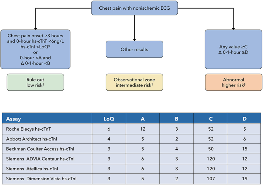
Reichlin et al77 developed an algorithm incorporating both baseline hs-cTnT values and changes in hs-cTnT between 0 and 1 hour to assign patients to rule out, rule in, or observation zones. This approach has been replicated with multiple hs-cTnI assays and externally validated.76,78–80 This algorithm has been combined with the 0-hour rule out described previously, such that patients may be ruled out either by having a very low hs-cTn at baseline (if chest pain onset is ≥3 hours) or by having values below a specified threshold and no more than a very small change (”delta”) between the serial measurements (see Figure 3). This combined 0- and 0/1-hour pathway has been labeled the “ESC 0/1 algorithm.” Options are also available for 0/2-hour algorithms that are conceptually identical but may be logistically easier to implement in EDs that cannot consistently capture a second hs-cTn value 60 minutes after the first measurement (see Figure B in the Supplemental Appendix for 0/2-hour CDPs for ruling out MI). Because the change thresholds for the 0/1 and 0/2 algorithms are time-dependent, it is critical that the blood specimens are collected within the specified windows, and accurate timing of specimen collection should be a performance metric that is tracked by health care systems using CDPs. Specimens collected outside of the specific time window should be interpreted with consideration of the actual time the specimen was collected. Importantly, thresholds tested are assay-specific (see Figure 3 and Figure B in the Supplemental Appendix). Although the vast majority of studies evaluating the 0/1- and 0/2-hour protocols have been observational, several prospective studies and randomized trials where the 0/1-hour protocol has been used for patient care have found a 30-day death/MI rate of <1%.68,86,87
FIGURE 3. Modified European Society of Cardiology 0/1-Hour CDP for Ruling Out MI.
Note that variations of these rapid CDPs have been implemented in different centers, and modification of the algorithms shown may be considered based on local considerations. All values in the chart in are ng/L. *The LoQ may differ slightly from the 0-hour rule-out threshold tested in individual studies. Using a cutoff of <5 ng/L can also be considered instead of the LoQ for the 0-hour rule-out threshold for hs-cTnI assays. †See Sections 5.6 and 5.8 for recommendations on follow-up and testing. ‡See Section 5.5.3. Additional evaluation should include at least one additional observation with hs-cTn measurement at 3–6 hours, with classification of myocardial injury, as described in Section 5.7, into chronic myocardial injury, acute myocardial injury, type 1 MI, and type 2 MI, as per the Universal Definition of Myocardial Infarction. §Patients with acute MI should be managed according to standard practice guidelines.
Efficacy with these algorithms is typically defined as the proportion of patients ruled out for MI, with safety defined by sensitivity and NPV for MI diagnosis at the index presentation and freedom from death/MI at 30 days. Efficacy with the 0/1- and 0/2-hour algorithms is approximately 60%, with some variation due to different assay thresholds and populations tested. Safety is high, with sensitivities of ~99% and NPVs >99.5% for MI at the index admission demonstrated in a meta-analysis.88 These protocols rule out approximately 60% to 65% of individuals, “rule in” ~15% of individuals, and assign ~25% to 30% to an intermediate-risk/observation zone (see Section 5.5). Direct comparisons with the 0/3-hour hs-cTn protocol demonstrate that the 0/1-hour protocol rules out more patients (64% vs 49%; P < 0.001) with a similar safety profile.89 In a randomized controlled trial using hs-cTnT, an ESC 0/1-hour algorithm resulted in more frequent discharge from the ED (45% vs 32%; P < 0.001) and a 1-hour shorter ED length of stay (P < 0.001), with similar clinical outcomes at 30 days (P = 0.001 for noninferiority) compared with a modified usual care approach using 0/3-hour cTn measurements with an hs-cTnT threshold of ≥30 ng/L.68
In initial studies, the positive predictive value (PPV) for those assigned to the rule-in group was approximately 75%,77 higher than the 50% PPV seen when the 99th percentile threshold was used for rule-in.90 However, the rate of MI in the European cohorts where the algorithms were developed was high (>15%). When similar approaches have been applied to U.S. populations where cTn testing is used more liberally, the proportion with adjudicated MI is much smaller (in some centers <5%), and thus the PPV of these algorithms has been considerably lower, ranging from 20% to 50% in less-selected populations of ED patients. In contrast, the PPV was approximately 70% in a U.S. study that enrolled patients more carefully selected based on a higher probability of ACS, and also incorporated a higher “rule-in” cTn threshold of 120 ng/L.85
Given the low PPV for MI when these CDPs are used broadly (as is typical for U.S. EDs), we recommend avoiding the term rule in for these algorithms and instead classifying this group as “abnormal.” The term rule in should be reserved for the subset of patients in the abnormal group meeting Universal Definition of MI criteria for MI (see Section 5.7).14
The ESC 0/1- or 0/2-hour algorithms take advantage of the better sensitivity and precision of the assay to safely rule out a majority of patients presenting with possible ACS and avoid the issues inherent with differing 99th percentile thresholds, including sex and age influences. Although efficacy (the proportion of patients ruled out) differs across multiple subgroups, with lower proportions of men, the elderly, and those with diabetes or chronic kidney disease ruled out, safety remains high in all subgroups.91–93
Disadvantages of these protocols include algorithm complexity, sensitivity to timing of blood draws, and the relegation of approximately one-quarter of patients to an observation zone in which there is limited evidence available to guide subsequent evaluation and treatment (see Section 5.5.3 and Table 3). A modification of the 0/1-hour protocol has been proposed, adding a 3-hour measurement of hs-cTn for those in the observation zone and classifying all patients at 3 hours into rule out or abnormal based on hs-cTn changes through 3 hours,94,95 although a recent study has questioned whether this provides a sufficiently high NPV.96
CAD = coronary artery disease; EDACS = Emergency Department Assessment of Chest Pain; HEART = History, ECG, Age, Risk Factors, and Troponin; LBBB = left bundle branch block; LVH = left ventricular hypertrophy; RBBB = right bundle branch block.
An additional limitation of these algorithms is that they may be susceptible to missing MI among late presenters who are on a flat portion of a descending cTn trend, where little or no change may be evident over 1 to 2 hours.97 Identification of these patients requires correlation of the clinical presentation with hs-cTn results and consideration for additional serial hs-cTn measurements.
5.4.3. High-STEACS 0/3-Hour Algorithm
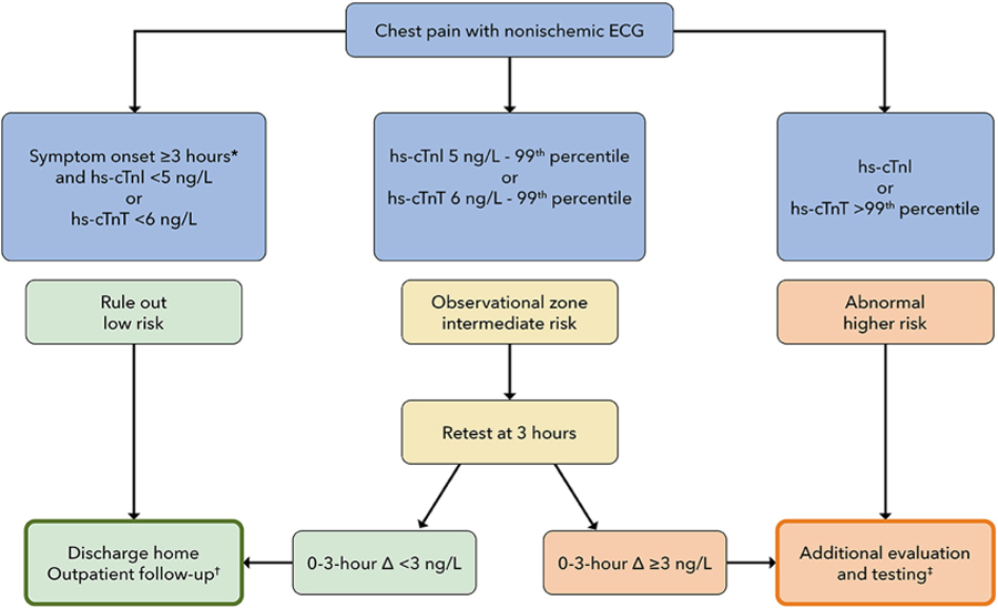
The High-STEACS algorithm (see Figure 4) is another validated implementation approach for hs-cTn. With this algorithm, MI is ruled out if the initial hs-cTnI is <5 ng/L; if hs-cTnI is ≥5 ng/L (or the patient is an early presenter) but hs-cTnI is less than the sex-specific 99th percentile URL, a second hs-cTnI measurement is performed 3 hours from the time of presentation. If the change from the first measurement is <3 ng/L and the value remains below the sex-specific 99th percentile URL, MI is ruled out. Although early presenters may be defined as those presenting within 2 hours of chest pain onset, we recommend using the more conservative 3-hour criterion for the time from chest pain onset, as described earlier. The High-STEACS algorithm was studied in a stepped-wedge randomized implementation trial in the EDs of 7 hospitals in Scotland (N = 31,492 individuals).61 Implementation of the pathway was associated with a reduction in length of stay from 10.1 ± 4.1 to 6.8 ± 3.9 hours (P < 0.001) and an increase in the proportion of patients discharged from the ED from 50% to 71%, with similar 30-day safety outcomes preimplementation and postimplementation. It is important to note that in the preimplementation period, the usual care protocol performed measurements of hs-cTnI at 0 and 6 to 12 hours, which is more conservative than “usual care” in most U.S. hospitals and may exaggerate benefits on length of stay vs what would be expected with implementation in the United States. Nevertheless, the High-STEACS pathway represents a validated approach for rule out that capitalizes on hs-cTn assay strengths. Observational data from the United States have demonstrated a similar safety profile with 100% sensitivity and 100% NPV for 30-day death or MI when combined with a normal ECG.62 In a subsequent analysis of the High-STEACS trial, the investigators demonstrated that the algorithm also performed well with the Roche fifth-generation hs-cTnT assay using the LoQ cutoff of 6 ng/L.75
*Although clinical trials with the HIGH-STEACS pathway have required chest pain onset ≥2 hours before presentation, we recommend requiring a ≥3-hour period between chest pain onset and the first troponin measurement in order to qualify for rule out at time 0. †See Sections 5.6 and 5.8 for recommendations on outpatient follow-up and testing. ‡Additional evaluation is recommended, with consideration of hospital observation or admission, and noninvasive anatomical or functional testing as described in Section 5.6. Myocardial injury should be classified as described in Section 5.7 into chronic myocardial injury, acute myocardial injury, type 1 MI, and type 2 MI, as per the Universal Definition of Myocardial Infarction. Patients with acute MI should be managed according to standard practice guidelines. Patients with chronic myocardial injury may be appropriate for discharge and management in an outpatient setting (see Sections 5.7 and 5.8).
ACS = acute coronary syndrome; ECG = electrocardiogram; High-STEACS = High-Sensitivity Troponin in the Evaluation of Patients With Acute Coronary Syndrome; hs-cTnI = high-sensitivity cardiac troponin I; hs-cTnT = high-sensitivity cardiac troponin T; MI = myocardial infarction.