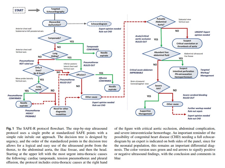
Here is an article on the SAFE-R Protocol, “Playing it SAFE in the NICU” SAFE-R: a targeted diagnostic ultrasound protocol for the suddenly decompensating infant in the NICU. [PubMed Abstract] [Full-Text HTML] [Full-Text PDF]. Eur J Pediatr. 2022 Jan;181(1):393-398. doi: 10.1007/s00431-021-04186-w. Epub 2021 Jul 5.
Today, I review, link to, and embed NeoPocusCollab‘s Targeted Use of POCUS for a Crashing Infant in the NICU: SAFE-R Protocol.
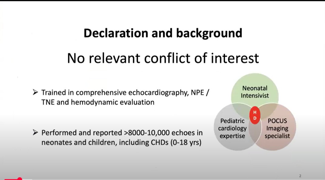
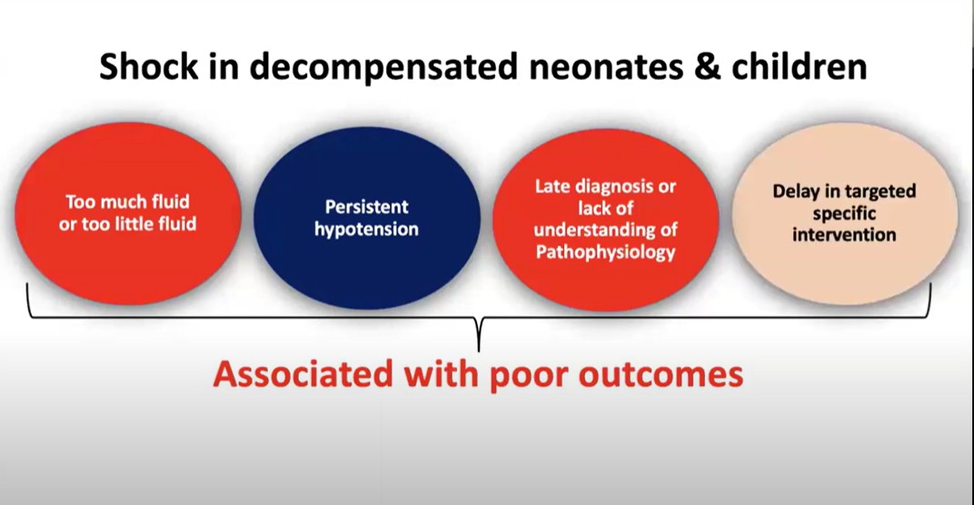
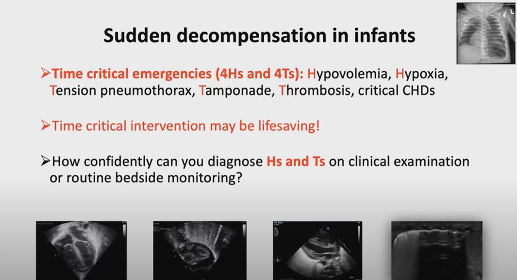
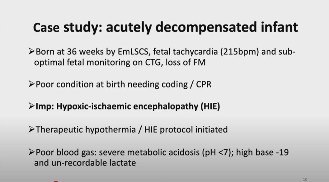
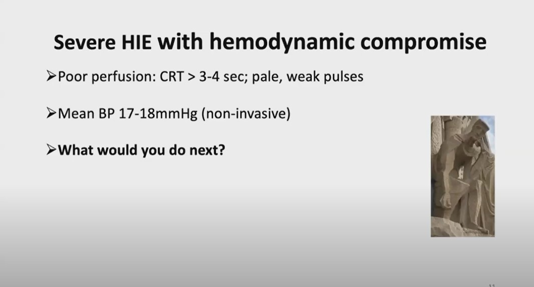
All that follows is from the above resource.
Oct 23, 2024Dr. Yogen Singh discusses the SAFE-R Protocol for POCUS in a Crashing Infant in the NICU in the latest NPC Webinar
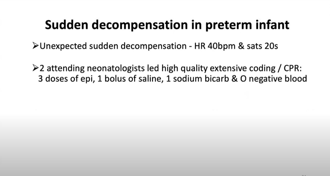
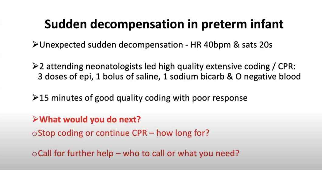
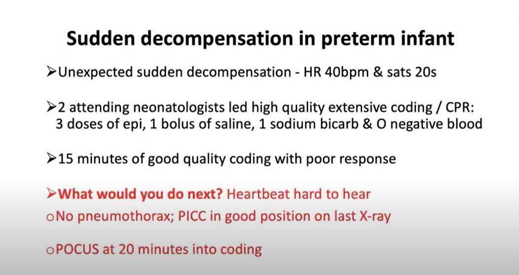
2:14
2:30
2:43
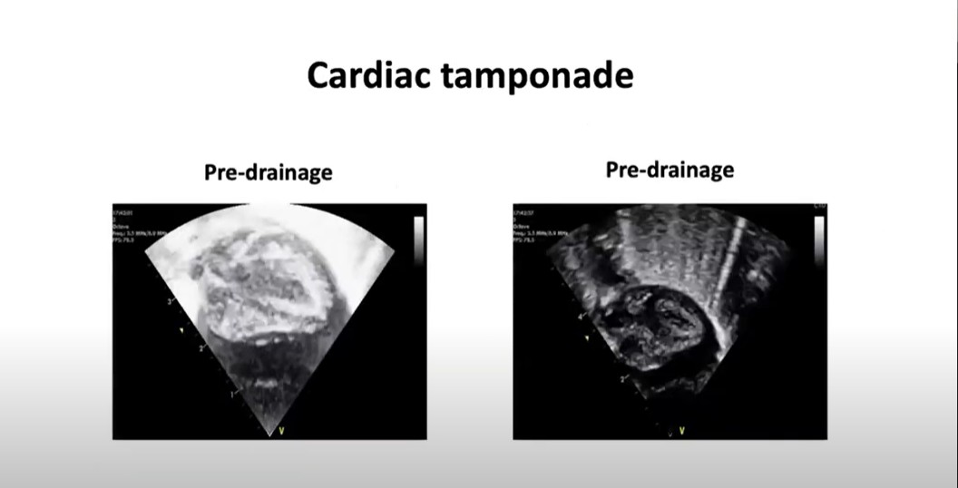
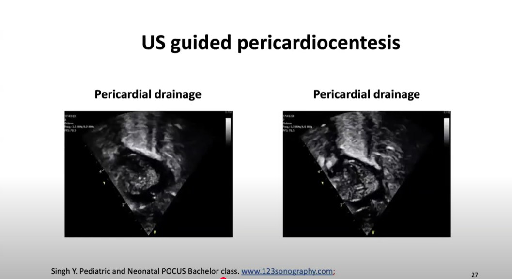
5:05
5:20
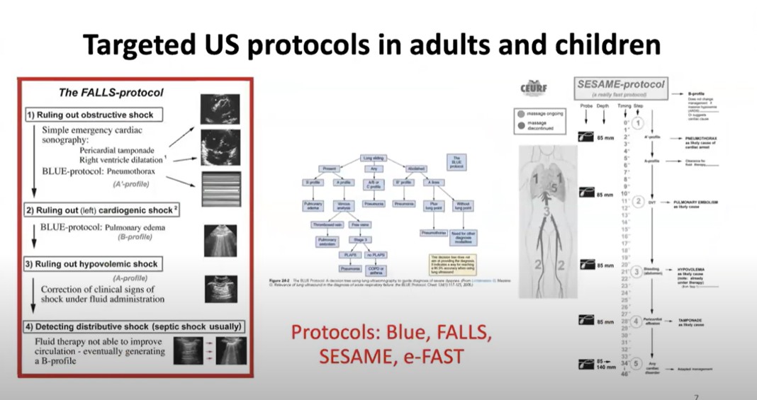
Lung ultrasound for causal diagnosis of shock (FALLS-protocol), a tool helping to guide fluid therapy while approaching fluid tolerance. Some comments on its accuracy [No abstract available] [Full-Text HTML] [Full-Text PDF]. Daniel A. Lichtenstein1* and Stéphane Bar2. Ann Intensive Care. 2024 Jun 14;14(1):88. doi: 10.1186/s13613-024-01329-8.
Acute respiratory failure: The BLUE protocol
May 11, 2023 NYSORA
The SESAME Protocol for the POCUS assessment of the etiology of cardiac arrest.
FAST Scan-eFAST Ultrasound Exam Made Easy: Step-By-Step Guide by POCUS 101.
6:26
7:32
8:30
8:43
9:00
9:42
10:25
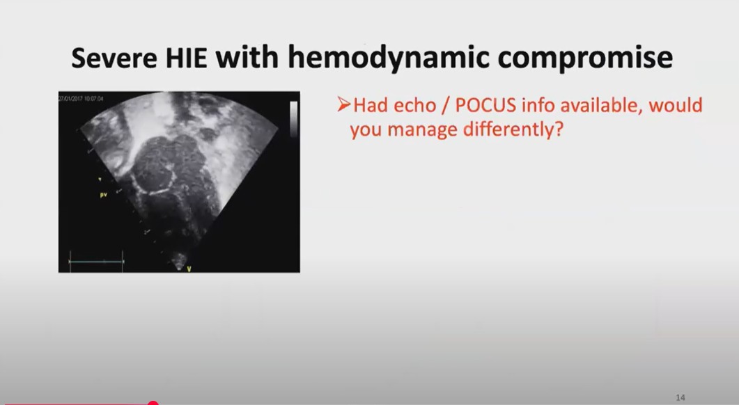
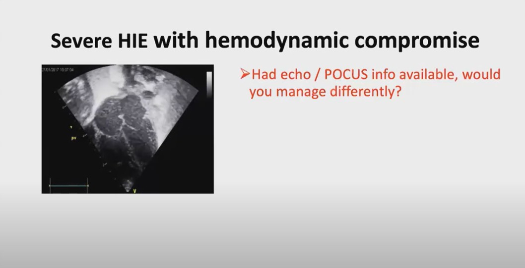
10:41 The same baby but it comes from an outlying hospital which has performed a pocus study which is available. This is an apical fourchamber
11:25
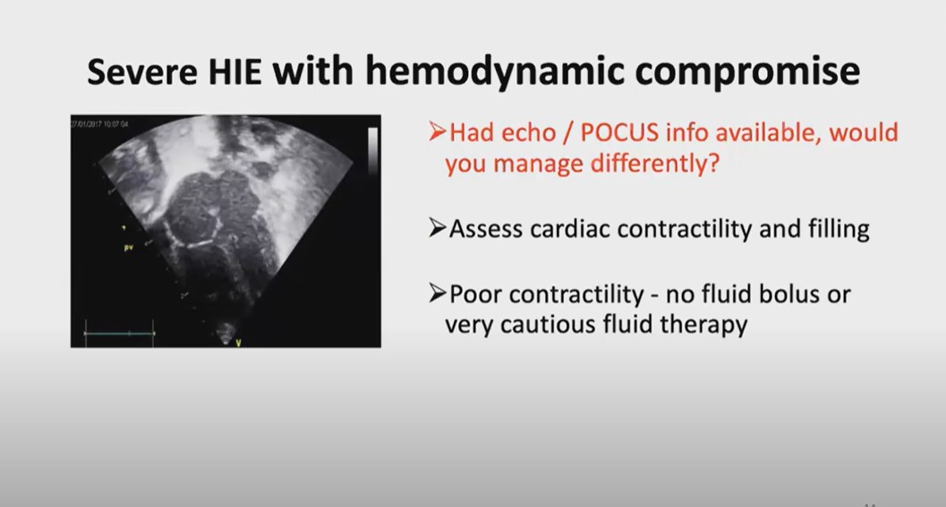
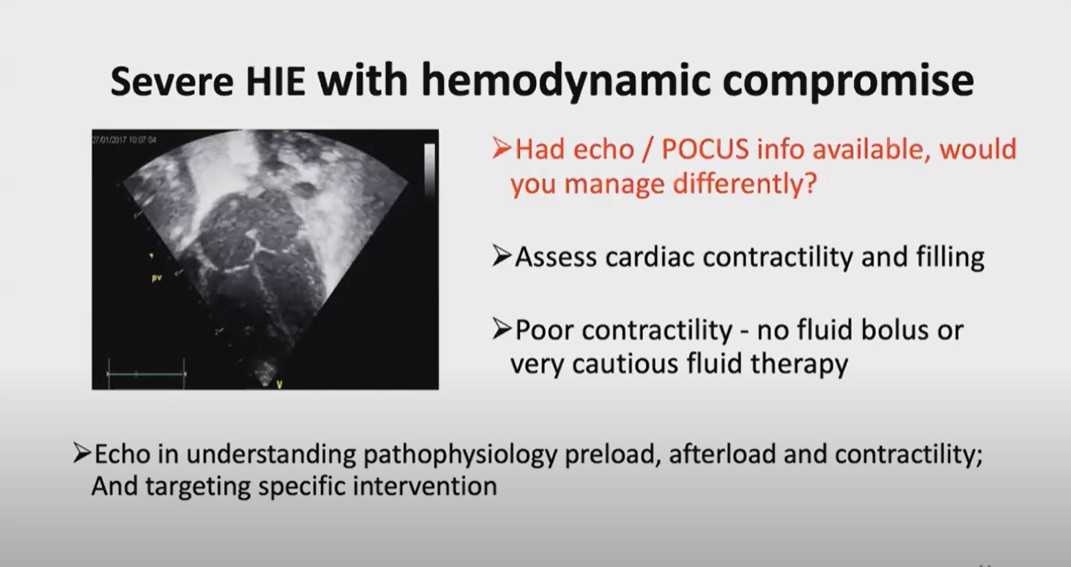
the same baby now comes from the local community hospital to the tary unit after a few hours about after about
four hours someone does a very limited Focus that point it’s not the best image
but despite not the best quality image is what do you see is apical four
chamber View you can see very poor card contac and heart is well filled there’s
no signs of hypovolemia heart is the cavities are B in look on the left
ventricle you can see really nice and open cavity here and this the left ventricle here left atrium right atrium
right ventrical just see this simple image would have been available on the bed
side just ask yourself will you get a fuel buus now and answer simple answer
no so very Target Focus can provide in if emergency situations very important
information it will not tell about the cardic functions what percentage of ejection fraction is but for the
Emergency lifethreatening it will tell the contact lady tell whether you should get fluid bolus or avoid the fluid bolus
course youve got the expertise for the hamic evaluation T you can look on the cardiac outputs and more
detailed hemic evaluation which can can be even more helpful let’s look on the second case
The Second Case:
11:33
11:52
12:04 Second Case:
Persistent Pulmonary Hypertension of the Newborn from StatPearls. Last Update: July 31, 2023. Padma S. Nandula; Sanket D. Shah.
13:21
on ECMO watch-Patients with the potential to be placed on ECMO.*
*See Neonatal extra corporeal membrane oxygenation, Indian J Thorac Cardiovasc Surg. 2020 Sep 3;37(4):411–420. doi: 10.1007/s12055-020-01005-z. There are 96 similar articles in PubMed.
13:48
14:02
15:02
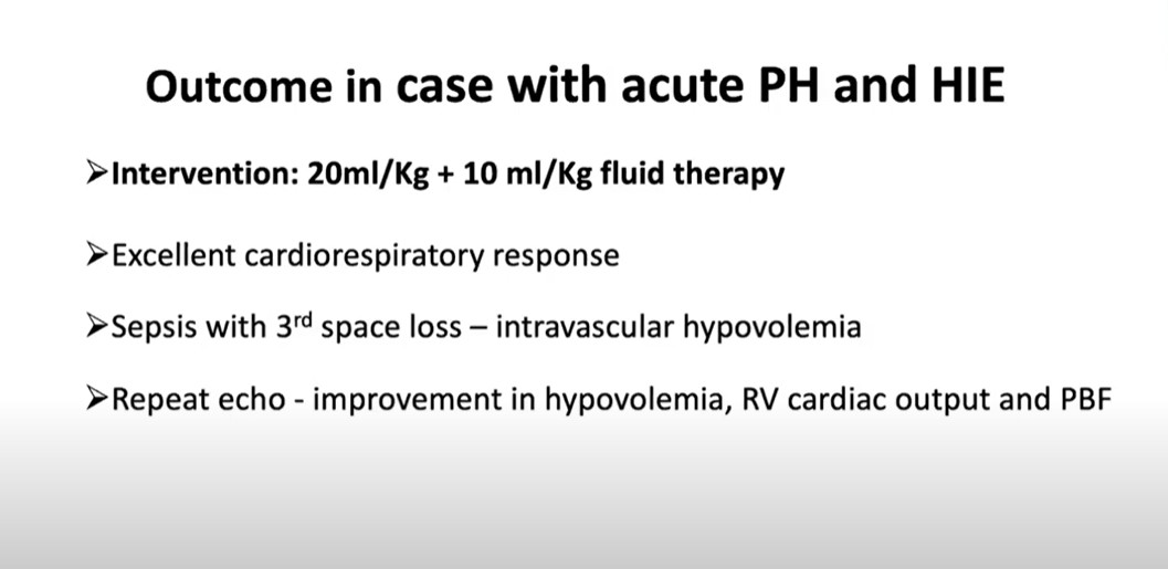
I’ll present only the
pocus part today and in this case how that can be helpful even let’s look on these two
views on this side you can see the epical four chamber V and you can see the rights of the heart is much bigger
right ventricle is almost double the size maybe bigger than the right left ventricle see with the right atrium is
much bigger in the par long AIS V right
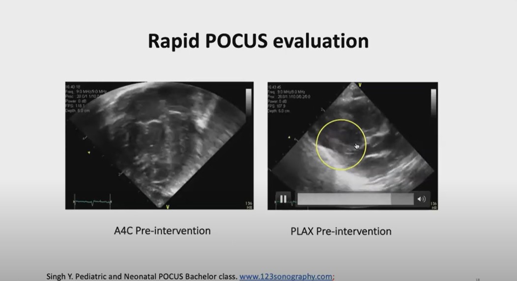
ventrical is is big is Hy trage in vental septum is here and you can see
the poster free wall which is left vental freeall here but you can see in the para
long access V the in vental septum and the poster free wall they’re touching
each other we call it a kissing sign of the left ventricle that’s a good sign of the
hypovolemia sometime with a bad like significantly hyper of left right ventricle in ventrical sep may come
towards the Left Post wall but hardly touches but here you can see it’s almost
dividing the cavity into two one towards the Apex once toward the left ventrical
outl track here just in the focus on the focus
images you can look on the multiple views maybe at least three views epical four chamber per perest long ACC View
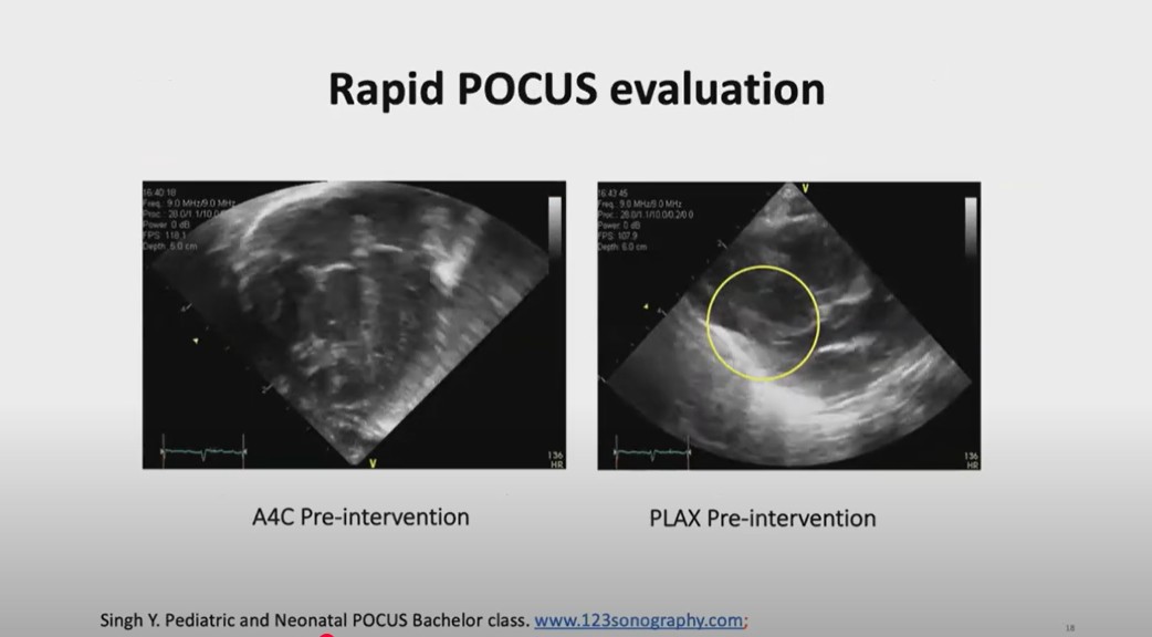
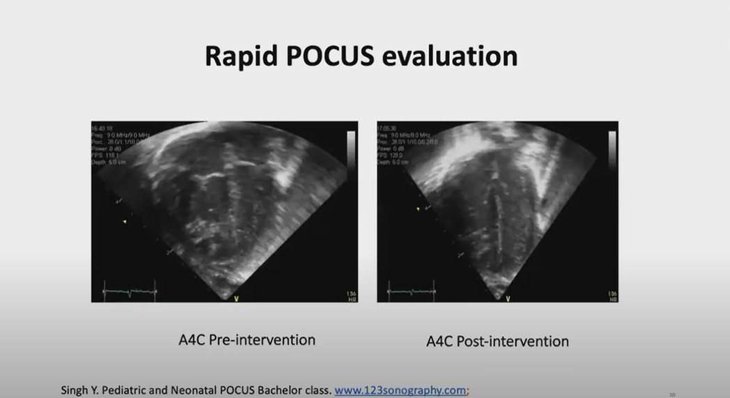
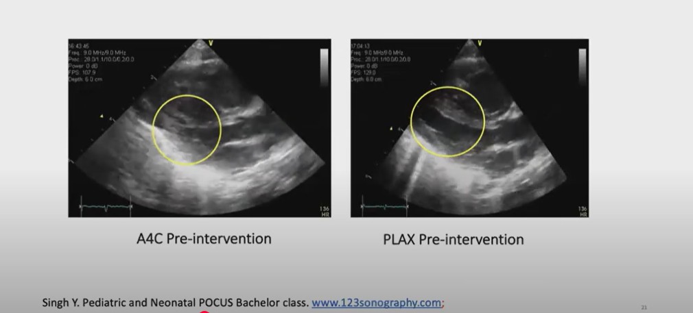
15:2716:2716:41 [These images below are mislabeled, I think. Left image is PLAX pre-intervention. Right image is PLAX post intervention. Left image shows post free wall and septum kissing. Right image shows post intervention and the left ventricular volume has increased.]
18:52
19:00 Third Case:20:1821:10
21:3322:40