
Today, I review, link to, and excerpt from MDPI Life‘s A Narrative Review of Point of Care Ultrasound Assessment of the Optic Nerve in Emergency Medicine. [PubMed Abstract] [Full-Text HTML] [Full-Text PDF]. Life (Basel). 2023 Feb 15;13(2):531. doi: 10.3390/life13020531.
There are 104 similar articles in PubMed.
The above article has been cited by 8 articles in PubMed.
All that follows is from the above article.
Abstract
Point of care ultrasound (POCUS) of the optic nerve is easy to learn and has great diagnostic potential. Within emergency medicine, research has primarily focused on its use for the assessment of increased intracranial pressure, but many other applications exist, though the literature is heterogeneous and largely observational. This narrative review describes the principles of POCUS of the optic nerve including anatomy and scanning technique, as well as a summary of its best studied clinical applications of relevance in emergency medicine: increased intracranial pressure, idiopathic intracranial hypertension, optic neuritis, acute mountain sickness, and pediatric intracranial pressure assessment. In many of these applications, sonographic optic nerve sheath diameter (ONSD) has moderately high sensitivity and specificity, but the supporting studies are heterogeneous. Further studies should focus on standardization of the measurement of ONSD, establishment of consistent diagnostic thresholds for elevated intracranial pressure, and automation of ONSD measurement.
Keywords: acute mountain sickness; idiopathic intracranial hypertension; intracranial pressure; optic nerve; optic nerve sheath diameter (ONSD); optic neuritis; point of care ultrasound (POCUS); ultrasound.
4.2. Indications and Contraindications
EPs working in a busy department will most effectively use POCUS to answer specific clinical questions to guide further management of patients. The primary question is whether there is sonographic evidence of elevated ICP. As such, clinical indications generally revolve around the suspicion of raised ICP, including presentations of head trauma, headache, stroke, or altered level of consciousness [2]. In trauma patients with periorbital swelling, POCUS allows ocular assessment for elevated ICP without requiring eyelid retractors or sedation to examine for papilledema. Furthermore, POCUS of the optic nerve may be useful for assessing and monitoring ICP in patients who are sedated and paralyzed, such as the intubated patient, where neurological examination is unreliable. Other indications relate to the inability to access advanced neuroimaging or lack of specialist support, such as in rural or remote settings, or in patients that are too unstable to leave a monitored setting for advanced imaging [38].The main contraindication to POCUS of the eye and optic nerve is suspected or confirmed globe rupture, but this may not be apparent to the clinician due to periorbital swelling or hematoma. Therefore, it is imperative to use plenty of ultrasound gel and to avoid direct ocular pressure while scanning in the setting of trauma [39]. Another relative contraindication is uncontrolled open angle or acute angle glaucoma [2].4.3. Scanning Technique and Sonographic Anatomy
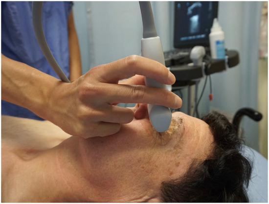
POCUS of the optic nerve is performed using a high-frequency linear transducer (5–14 MHz) on an ultrasound machine, with a smaller probe footprint providing better sonographic contact with the eye [33,40]. An ocular preset should be used, limiting thermal index to ≤1 and mechanical index to ≤0.23 in accordance with the as low as reasonably achievable (ALARA) principle [39,41]. Patients are placed in a comfortable position, generally supine, and asked to close their eyes with their head in a neutral position (Figure 2) [42]. Copious sterile ultrasound gel should be placed on the closed eyelids of the patient, and the probe should be placed gently on top of the gel to avoid direct pressure on the eye. The sonographer’s hand can be steadied over the patient’s zygomatic process or nasal bone to regulate the amount of downward pressure applied by the probe. If preferred, a transparent protective film may be placed over the eye to protect it, but care must be taken to avoid air entrapment between the film and external eyelids, as this will degrade image quality [41]. Scanning should be performed using B-mode in the transverse orientation. The patient should be asked to look forward at a fixed point in neutral gaze to minimize eye movement [2].Figure 2. Demonstration of point of care ultrasound of the eye. Plenty of ultrasound gel should be used and the hand can be steadied over the patient’s zygomatic process to minimize downward pressure on the eye applied by the probe. A clear, protectant film may be used to protect the eye if desired. This image was obtained with consent from the model for publication.The optic nerve can be identified as a homogeneous hypoechoic linear structure extending far-field from the anechoic ocular globe [41]. It is surrounded by a more hyperechoic band representing the optic nerve sheath, analogous to the arachnoid membrane surrounding the subarachnoid space, which is further enclosed by a hypoechoic outer band representing the dura mater [2,43]. Finally, hyperechoic retrobulbar fat surrounds the optic nerve. The central retinal artery and vein run through the middle of the optic nerve, while the ophthalmic artery runs parallel adjacent to the optic nerve [44].Traditionally, ONSD is measured 3 mm behind the globe where the sheath is the most distensible and its measurements are reproducible [36,40]. Though there is recent evidence suggesting no difference in ONSD when measured between 3 and 5 mm posterior to the globe [45], most studies base their measurements at 3 mm [43]. The ONSD is measured between the outer hyperechoic borders of the subarachnoid space (Figure 3) [2,46]. If no hyperechoic stripe is visualized, the diameter is measured at the border of the hypoechoic optic nerve and the hyperechoic retrobulbar fat [32]. The measured diameter should be perpendicular to the longitudinal axis of the optic nerve [43]. Color doppler may aid visualization of a tortuous optic nerve by visualizing the midline central retinal artery and vein, allowing for accurate measurement of the ONSD perpendicular to the true course of the optic nerve [44,47]. However, the use of doppler must be minimized to avoid ocular injury [39]. Averaging 2–3 measurements in transverse or sagittal planes may minimize measurement errors [40,41]. Like other ultrasound applications, repeated measurements as clinically indicated may provide additional dynamic information [2].Figure 3. Ultrasound image of the eye and optic nerve. A: eyelid, B: cornea, C: anterior chamber, D: iris, E: vitreous chamber, F: retina, G: optic nerve with optic nerve sheath diameter of 3.8 mm, measured at 3 mm from the retina. This figure was obtained with consent from the model for publication.POCUS of the optic nerve can easily be learned by novice users with minimal ultrasound experience after a brief training session [48]. EPs can accurately measure ONSD with ultrasound when compared to ONSD measurements on CT by a radiologist [49], and generally have good inter-rater reliability regardless of level of ultrasound training, though some variations exist amongst residency-level physicians [38,50].4.4. Clinical Applications: Increased Intracranial Pressure
The most prominent clinical application of optic nerve POCUS is its diagnostic ability to detect elevated ICP. Kim et al. conducted a prospective observational study in 2021, comparing ultrasound assessment of ONSD with CT of the brain performed within 30 min in patients suspected of raised ICP in the ED [4]. From a total of 199 enrolled patients, 57 were found to have signs of raised ICP on CT scan. The median ONSD on ultrasound in the raised ICP group was significantly higher compared with the normal ICP group (5.7 mm vs. 4.3 mm, p < 0.001). These results confirm the findings of another prospective observational study from 2019 by Hanafi et al. where 62 trauma patients were compared with 50 healthy controls [5]. Of the 55 trauma patients with increased ICP on CT scan, sonographic ONSD was 6.06 mm in these patients compared to 4.02 mm in healthy controls.Ohle et al. published a systematic review and meta-analysis in 2015 comparing trials assessing sonographic ONSD to diagnose elevated ICP compared to CT as the reference standard [6]. They included 12 studies with 478 study subjects in ED and ICU settings and found that ONSD had a sensitivity of 96% (95% CI, 88–99%), specificity 92% (95% CI, 78–98%), diagnostic odds ratio (DOR) 319 (95% CI 79–1290), positive likelihood ratio (+LR) 12.5 (95% CI 4.2–37.5), and negative likelihood ratio (–LR) 0.05 (95% CI 0.02–0.14). The major limitation of this meta-analysis was the moderate-to-high heterogeneity of the included studies.Recognizing that invasive monitoring is more accurate than CT to detect raised ICP, Robba et al. performed a meta-analysis in 2018 that only included studies using invasive ICP measurement (intraparenchymal, intraventricular, or lumbar puncture [LP]) as the reference standard [7]. Raised ICP was defined as >20 mmHg or >25 cmH2O. They included seven studies with a total of 320 patients and found a pooled DOR of 68 (95% CI 29–135), +LR 5.4 (95% CI 3.8–7.5) and −LR 0.09 (95% CI 0.05–0.15). Their results are more modest compared to Ohle’s systematic review, but this likely relates to a more robust reference standard, fewer included studies, and lower heterogeneity. An updated meta-analysis by Aletreby et al. in 2022 assessed ONSD compared to invasive ICP measurement as the reference standard [8]. They included nine additional studies compared to Robba’s systematic review for a total of 619 patients. Their results were similar: pooled sensitivity of 90% (95% CI 85–94%), specificity 85% (95% CI 80–89%), DOR 47 (95% CI 26–83), +LR 6.1 (95% CI 4.4–8.5), and −LR 0.11 (95% CI 0.07–0.18).The largest meta-analysis of optic nerve POCUS was published by Koziarz et al. in 2019 and included trials with participants in all age groups, sonographers of any training level, and used any reference standard as the comparator [9]. Their analysis included 71 studies with a total of 4551 patients and found a sensitivity and specificity in the traumatic brain injury subgroup of 97% (95% CI 92–99%) and 86% (95% CI 74–93%), respectively. In non-traumatic brain injury, sensitivity was mildly reduced at 92% (95% CI 86–96%) while specificity was similar at 86% (95% CI 77–92%).Most studies used differing ONSD thresholds to diagnose increased ICP [4,8]. In 2019, Kim et al. attempted to establish a single sonographic ONSD cut point to detect elevated ICP [10]. They included six studies with 352 participants in their final analysis, all utilizing 5 mm as a cut point. They found this cut point provided a pooled sensitivity of 99% (95% CI 96–100%), specificity 73% (95% CI 65–80%), DOR 178 (95% CI 53–599), +LR 4.6 (95% CI 2.0–10.9), and −LR 0.05 (95% CI 0.02–0.14).In comparison, the physical exam is generally poorly sensitive and specific for elevated ICP. Fernando et al. compared multiple modalities to diagnose elevated ICP, including physical exam and sonographic ONSD. Their meta-analysis found pupillary dilation was insensitive at 28% but moderately specific at 86%. Motor posturing had poor sensitivity and specificity at 54% and 64%, respectively. Decreased level of consciousness was more sensitive at 76% but less specific at 40% [11]. In comparison, sonographic ONSD measurement had a pooled area under the receiver operating characteristic (ROC) curve of 0.94. These authors did not calculate pooled sensitivity and specificity as the included studies used many different optimal cut points for elevated ICP. Finally, while direct ophthalmoscopy can detect elevated ICP based on the presence of papilledema, it is infrequently and poorly performed by EPs [51].Based on the literature, optic nerve POCUS using an ONSD threshold of 5 mm appears to be a clinically useful and accurate tool to detect elevated ICP in the emergency setting. In general, it seems to be more sensitive than specific, which is clinically appropriate given that it would be undesirable to miss a case of high ICP. However, the lack of large randomized controlled studies limits the widespread use of this tool. POCUS assessment of ONSD cannot replace CT or invasive monitoring to diagnose elevated ICP, but it can be used in an appropriate clinical context to monitor at-risk patients, in patients too unstable to leave a monitored setting, or as a supplementary test in settings where access to advanced neuroimaging is limited.4.5. Clinical Applications: Idiopathic Intracranial Hypertension
IIH is a neurologic disorder characterized by diffuse headache, visual abnormalities, and papilledema, most commonly affecting young females with increased body mass index (BMI) [16]. Physicians who are not ophthalmologists may have difficulty performing ophthalmoscopy for a variety of reasons, including limited equipment, lack of experience, or patient factors [52]. Therefore, POCUS of the optic nerve can be an effective alternative means to assess for elevated ICP and assist with the diagnosis of IIH.Case-control studies have assessed the diagnostic accuracy of sonographic ONSD to detect elevated ICP to diagnose IIH, generally finding good sensitivity and specificity with varying diagnostic thresholds. Dağdelen et al. measured ONSD in 47 subjects with IIH and 50 healthy controls. They found the mean ONSD in IIH patients was increased at 6.4 mm compared with 4.9 mm in controls (p < 0.001). They identified an optimal cut point of 5.7 mm, yielding a sensitivity of 100% and specificity of 98% [12]. However, Kishk et al. found more modest results when they compared 99 females with both clinically definite and probable IIH to 35 healthy controls. All cases had both neurologic and ophthalmologic assessment including neuroimaging to rule out other diagnoses. ONSD measurement was performed prior to diagnostic LP. Mean sonographic ONSD was higher in cases than controls (6.57 mm vs. 5.50 mm, p < 0.001). Using a cut point of 6.05 mm, sonographic ONSD had only modest sensitivity but good specificity at 73% and 91%, respectively [13]. Their more modest results may be due to the inclusion of nine cases with probable IIH. Smaller studies have demonstrated good diagnostic utility for ONSD measurement. Del Saz-Saucedo et al. studied 30 subjects with suspected IIH, of which 19 were diagnosed with a positive LP. Using a higher cut point of 6.3 mm, sonographic ONSD had a sensitivity of 95% (95% CI 82–100%) and specificity of 91% (95% CI 69–100%) [14]. Finally, Ebraheim et al. studied 24 patients with suspected IIH, of which 20 were diagnosed with a positive LP. Sonographic ONSD had a sensitivity of 88% and specificity of 100% using a cut point of 6.2 mm [15].Multiple studies have demonstrated dynamic changes in sonographic ONSD after LP. Jeub et al. found that removal of 30 mL of CSF by LP led to a reduction in ONSD by 0.4 mm and 0.5 mm in the right and left eyes, respectively [16]. In another study, Del Sez-Saucedo found that ONSD decreased by a mean of 0.9 mm after therapeutic LP achieved a CSF pressure of <15 cmH2O [14]. Even without therapeutic LP, Ebraheim found that after 4 weeks of treatment with acetazolamide alone, ONSD decreased by a mean of 0.4 mm [15].ONSD appears to have good sensitivity and specificity to diagnose IIH. Further, it can be used to monitor the response to treatment. However, no standardized cut points currently exist, limiting its use as a diagnostic tool. Ultimately, more studies are required to establish a standard cut point to serve as a diagnostic threshold for IIH.4.6. Clinical Applications: Optic Neuritis
Optic neuritis is an acute inflammatory disorder of the eye that causes vision disturbance or loss and ocular pain. It is most commonly idiopathic but may be secondary to other disease states, most notably multiple sclerosis [30]. Diagnosis is largely clinical and is aided by MRI [30,53]. Numerous case reports describe increased ONSD when measured by POCUS in patients ewith optic neuritis, leading to observational studies to research its potential as a diagnostic tool [54,55,56].Lochner et al. studied 21 patients with first-episode demyelinating unilateral optic neuritis and 21 matched controls. All patients underwent MRI imaging to rule out other causes for their symptoms. They determined that sonographic ONSD was significantly increased compared to the unaffected eye, median 6.3 mm vs. 5.5 mm, respectively, and ONSD was larger in affected eyes than in controls. ONSD of the unaffected eye was similar to controls [17]. Kwon et al. studied 17 patients with new-onset unilateral optic neuritis and found median ONSD to be modestly higher in affected eyes than in unaffected eyes, measuring 5.51 mm and 5.05 mm, respectively [18]. While their findings are modest, they are supportive of the findings from Lochner’s study. As only two studies exist, more research is required to establish sonographic ONSD as a useful tool to diagnose optic neuritis.

See Optic Neuritis, StatPearls, Last Update: January 20, 2025.
4.7. Clinical Applications: Acute Mountain Sickness
Optic nerve sheath sonography has also been explored as a portable and non-invasive surrogate measure for increased ICP in acute altitude illnesses. Although causality is not clearly established, increased ICP has been reported in patients with AMS and high-altitude cerebral edema (HACE). Both entities often overlap with high altitude pulmonary edema (HAPE) [57,58].Further research is necessary to clearly establish whether there is a correlation between ONSD and AMS. Ideally, there needs to be a large longitudinal cohort study measuring ONSD at baseline followed by a range of altitudes at consistent time intervals, using a standardized scanning protocol with assessment of interobserver reliability, and controlling for confounding factors such as medication use (such as acetazolamide and dexamethasone), ascent time, and acclimatization periods.4.8. Clinical Applications: Pediatrics
There is particular interest in POCUS of the optic nerve in pediatric populations where radiation exposure for diagnostic imaging and invasive procedures are ideally avoided if reasonably possible. However, the measurement of ONSD in children is more complicated than in adults. ONSD increases with age, most rapidly in the first year of life [60]. Furthermore, ONSD may be affected by the patency of the anterior fontanelle and head circumference [27,41,61,62]. Because of these factors, no standardized ONSD normal value exists, though numerous diagnostic cut points for raised ICP have been suggested. One commonly cited ONSD cut point is 4 mm in children ≤1 year old, 4.5 mm in children 1 to 15 years old, and 5 mm in children >15 years old [41,60].4.8. Clinical Applications: Pediatrics
There is particular interest in POCUS of the optic nerve in pediatric populations where radiation exposure for diagnostic imaging and invasive procedures are ideally avoided if reasonably possible. However, the measurement of ONSD in children is more complicated than in adults. ONSD increases with age, most rapidly in the first year of life [60]. Furthermore, ONSD may be affected by the patency of the anterior fontanelle and head circumference [27,41,61,62]. Because of these factors, no standardized ONSD normal value exists, though numerous diagnostic cut points for raised ICP have been suggested. One commonly cited ONSD cut point is 4 mm in children ≤1 year old, 4.5 mm in children 1 to 15 years old, and 5 mm in children >15 years old [41,60].Şik et al. studied 147 children presenting to the ED with head trauma requiring a CT head. ONSD ultrasound was performed by a pediatric EM fellow who was blinded to clinical and radiologic findings. When compared to CT as the reference standard, they found a high sensitivity of 93% and specificity of 94% when using a cut point of 5.1 mm amongst all age groups [26]. However, others found sonographic ONSD to have lower diagnostic performance consistent with the results of Bhargava’s systematic review. Padayachy et al. performed a relatively large observational study comparing sonographic ONSD to invasive ICP measurements. They included 56 children ≤1 year old and 118 children >1 year of age. In children ≤1 year old using a cut point of 5.16 mm to detect ICP above 20 mmHg, sonographic ONSD had a sensitivity of 80% (95% CI 44–98%) and specificity of 76% (95% CI 61–87%), while in children >1 year, a cut point of 5.75 mm produced a sensitivity of 86% (95% CI 75–93%) and specificity of 70% (95% CI 56–82%). They found children with an open anterior fontanelle had the poorest correlation between sonographic ONSD measurements and the reference standard [27]. Ultimately, ONSD appears to have high sensitivity and only modest specificity to detect raised ICP in pediatric populations, but further research is required, especially to determine age-appropriate cut points.There has been increasing interest in utilizing sonographic ONSD to detect VP shunt failure. While VP shunt malfunction results in raised ICP, children often present with non-specific symptoms overlapping with benign childhood ailments [63]. While imaging is heavily relied upon to diagnose VP shunt failure, CT and MRI are insensitive and children often require repeat CT imaging, thereby raising the risk of malignancy due to cumulative radiation exposure [64]. Lin et al. studied 32 patients with suspected VP shunt failure and compared sonographic ONSD measured by pediatric EPs against neuroimaging and a neurosurgical clinical diagnosis as reference standards. When compared to neuroimaging, sonographic ONSD had a sensitivity of 60% (95% CI 23–88%) and specificity of 67% (95% CI 48–81%). When compared to neurosurgical opinion, sensitivity rose to 75% (95% CI 30–95%) and specificity to 68% (95% CI 49–82%) [28]. Hall et al. studied 39 encounters suspicious for VP shunt failure and compared sonographic ONSD by pediatric EPs with neurosurgical diagnosis.* They found sensitivity to be 61% (95% CI 36–83%) and specificity to be 22% (95% CI 6–48%) [29]. Unfortunately, sonographic ONSD appears to have poor diagnostic value in VP shunt failure, though larger studies in the future may provide clarity in this emerging area of interest.
*See Ventriculoperitoneal Shunt, StatPearls, Last Update: August 23, 2023.
Evaluation
Patients with shunts are evaluated for evidence of signs or symptoms related to complications or malfunction. Acute symptoms of malfunction/infection are: headache, lethargy, diplopia, nausea and vomiting, seizure, irritability, poor feeding, head enlargement when sutures are open, tense fontanelle, fever, neck rigidity. Shunt system has to be assessed manually for proper function and visible for evidence of redness or swelling along the shunt tubing. Shunt X rays are done to evaluate the integrity of the system. CT scan or MRI is done to evaluate the size of the ventricles.
In those cases of malfunction, the shunt has to be assessed for proper function. The following technique is used:
Complications of a shunt tap are:
Concluding today’s article:
5. Conclusions
POCUS of the optic nerve appears to be a useful tool for several clinical applications, including traumatic brain injury, IIH, and optic neuritis. Though evidence for its use is largely observational, sonographic ONSD generally has high sensitivity and moderate specificity to diagnose conditions that have a pathophysiologic process resulting in elevated ICP. It may also detect evidence of optic nerve inflammation in acute optic neuritis. While POCUS of the optic nerve should generally complement traditional diagnostic algorithms and modalities until larger randomized trials exist, it may be an important decision-making tool in specific circumstances such as the unstable patient or in rural or remote settings. Future studies should prioritize standardizing ONSD measurement techniques and diagnostic cut points in both pediatric and adult populations.


