In addition to the resource below, please review StatPearls‘ Abdominal Aortic Aneurysm Imaging. Last Update: November 28, 2022.
In this post, I link to and embed POCUS of the Aorta From MetroHealth Emergency Ultrasound, 45:47, Mar 16, 2023.
All that follows is from the above resource.
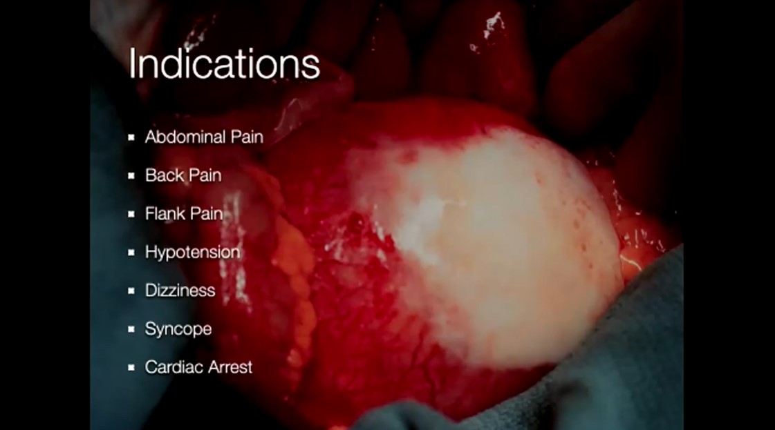
Indications for An Abdominal Aorta Scan
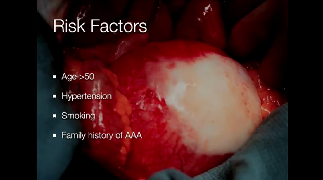
Risk Factors For Abdominal Aortic Aneurysm
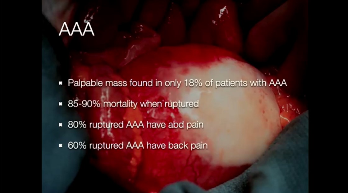
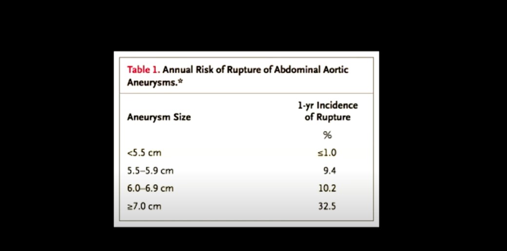
An Abdominal Aortic Aneurysm is greater than 3 cm.
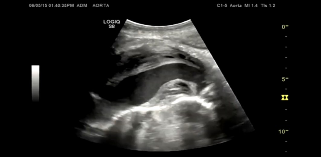
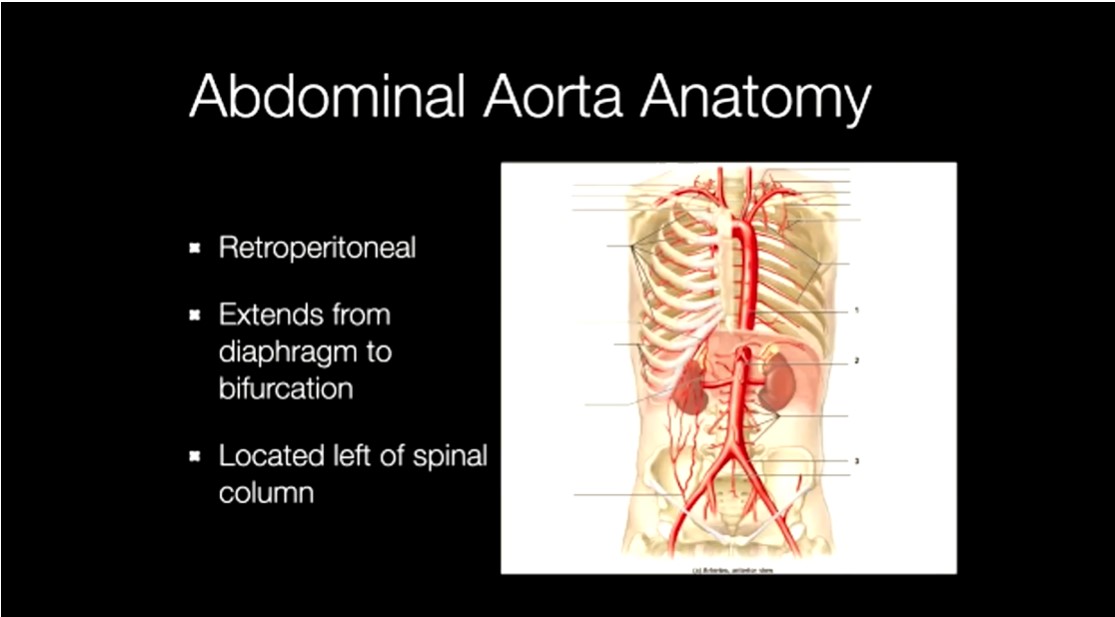
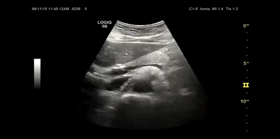
Long Axis of the Aorta
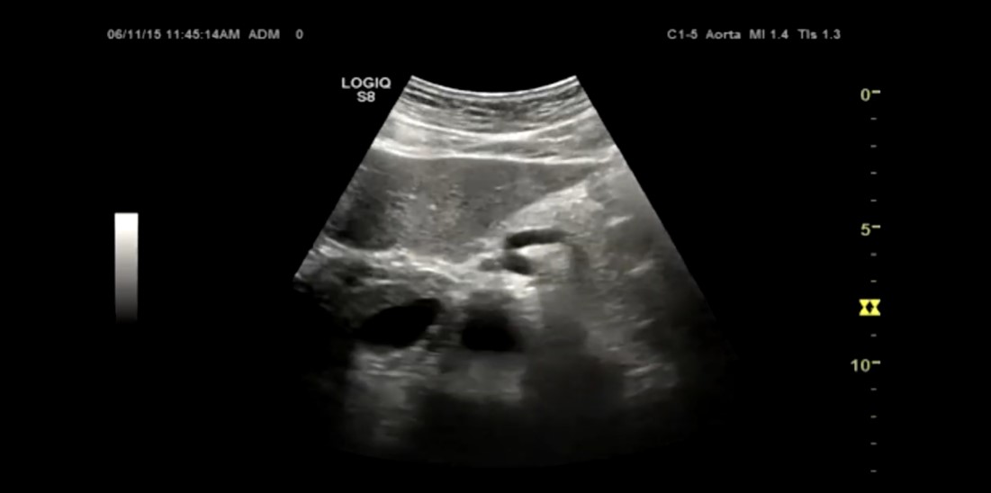
Bifurcation of the Aorta
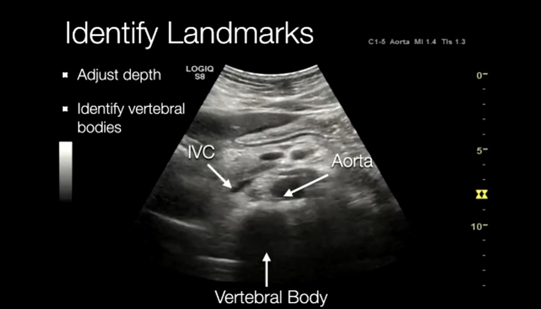
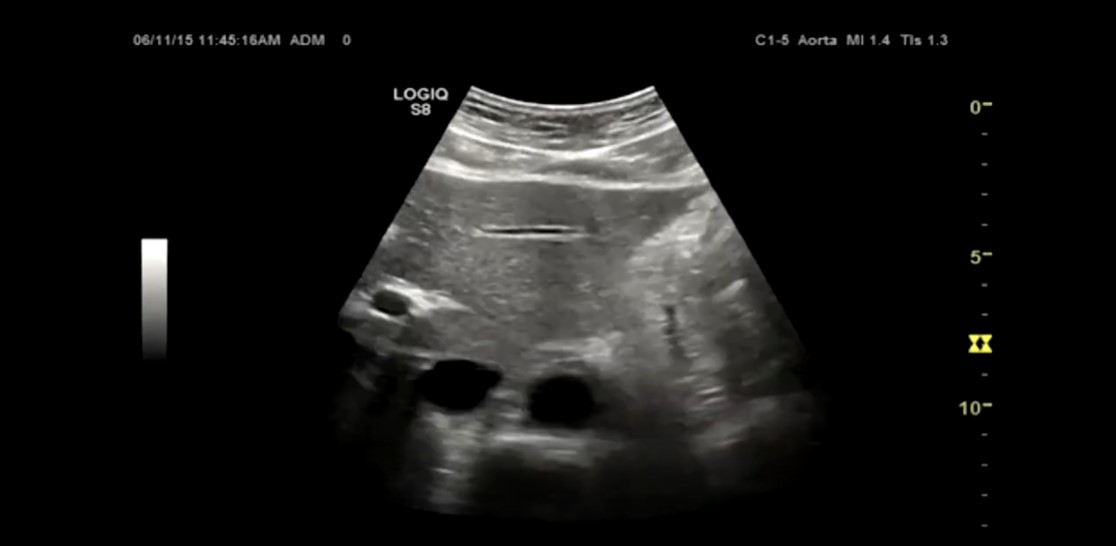
Short Axis of the Aorta in the center and the Inferior Vena Cava to the left of the Aorta on the screen
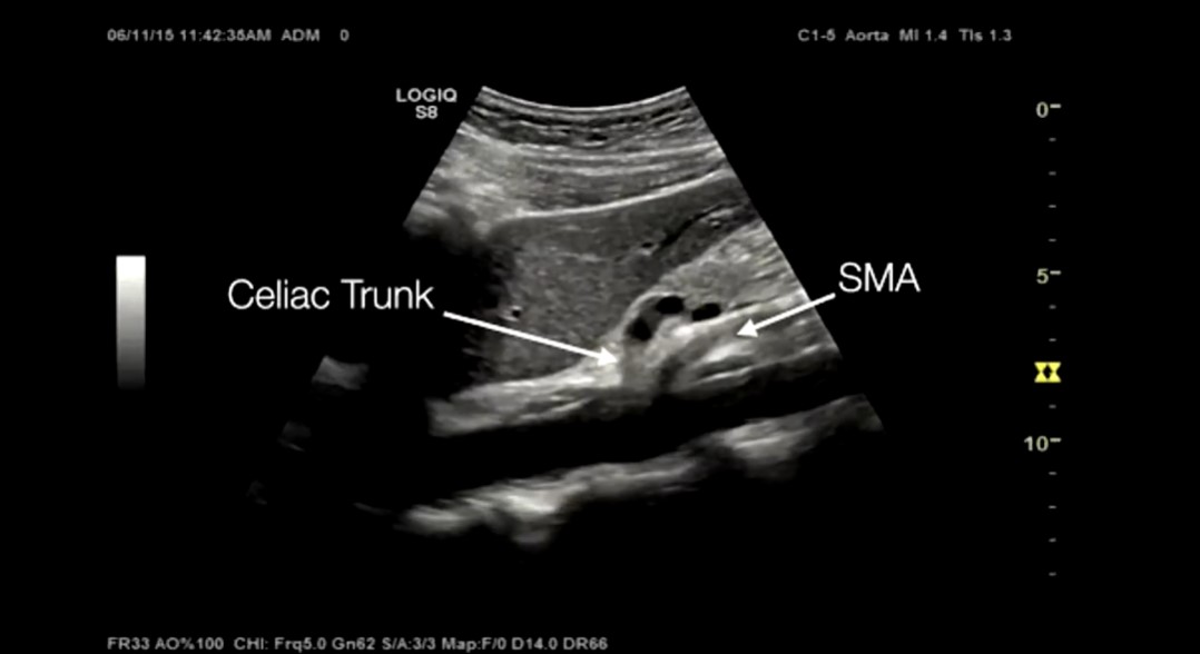
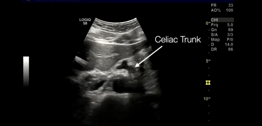
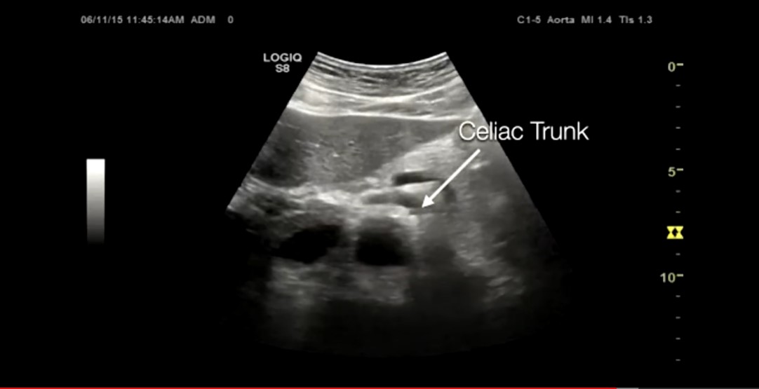
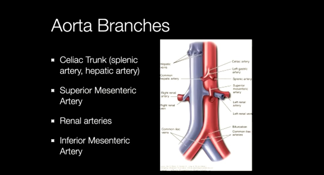
Celiac Trunk coming off of the aorta to the right of the screen (pt’s left) at one o’clock
Celiac trunk dividing into the splenic artery and the common hepatic artery. So this is the proximal view of the aorta.
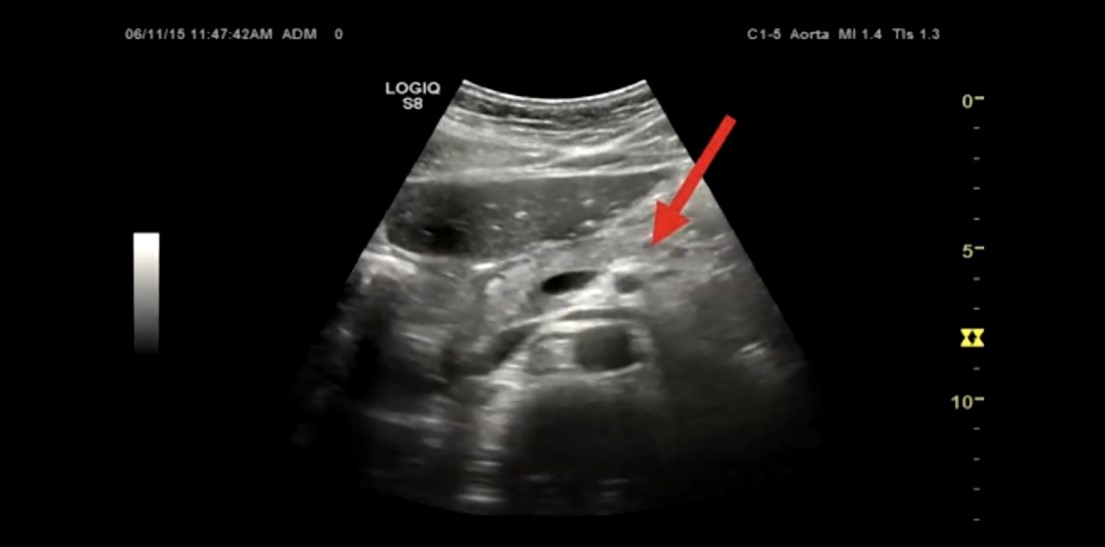
A little more distally on the aorta, we see the superior mesenteric artery (arrow) and the left renal vein coming off the IVC and the right renal artery coming of the aorta.
Above the superior mesenteric artery, is the splenic vein. And below the SMA above the aorta is the left renal vein.
Distal to the SMA, you won’t generally see the Inferior Mesenteric Artery.
And next, we see the aorta bifurcating into the Iliacs.
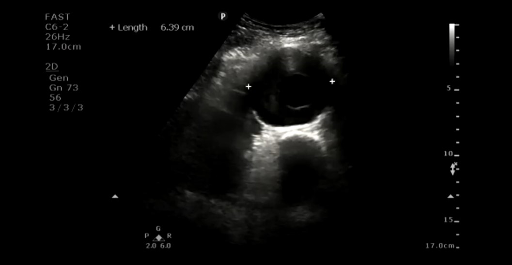
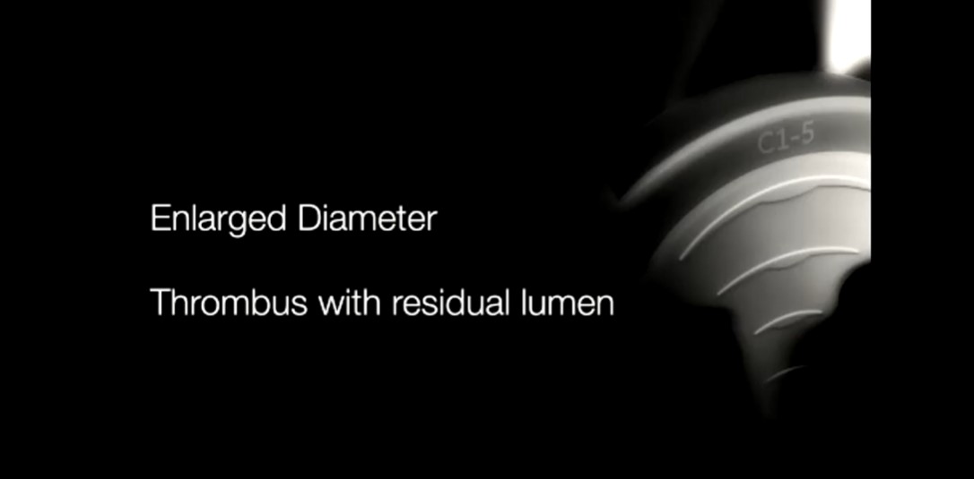
We measure the aorta from outer edge to outer edge.
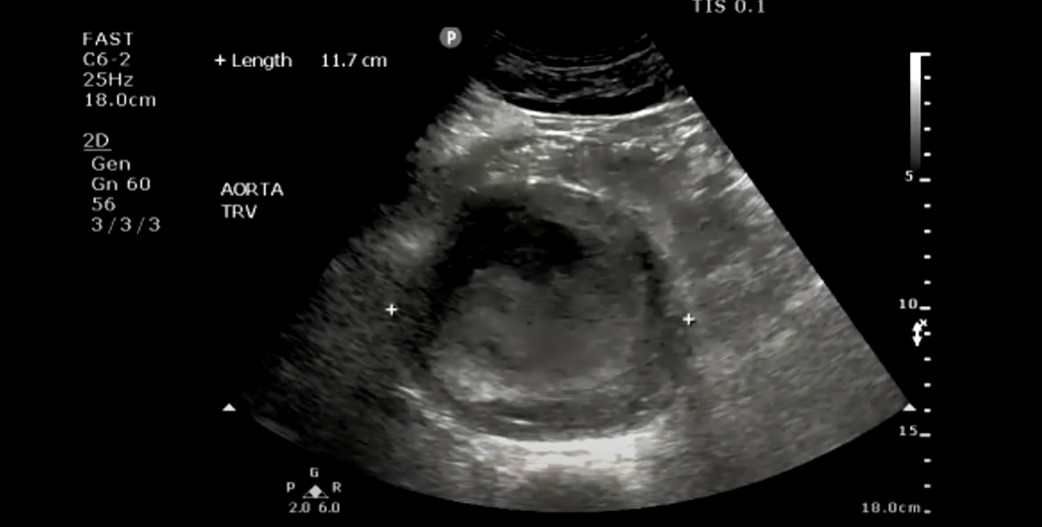
Note the central lumen with surrounding thrombus.
And here is the long axis. Note the large thrombus.
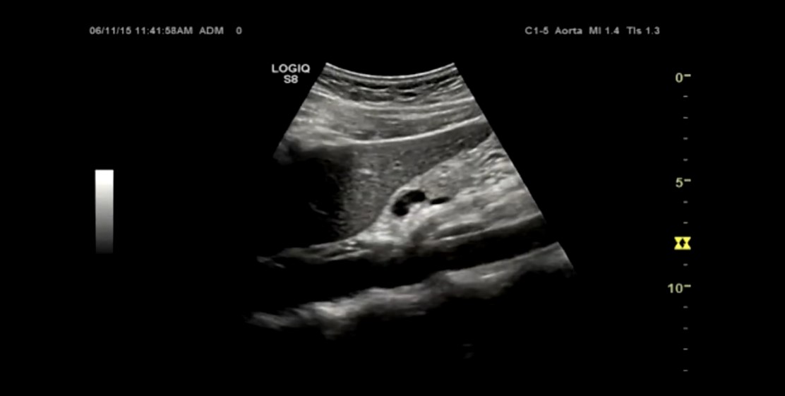
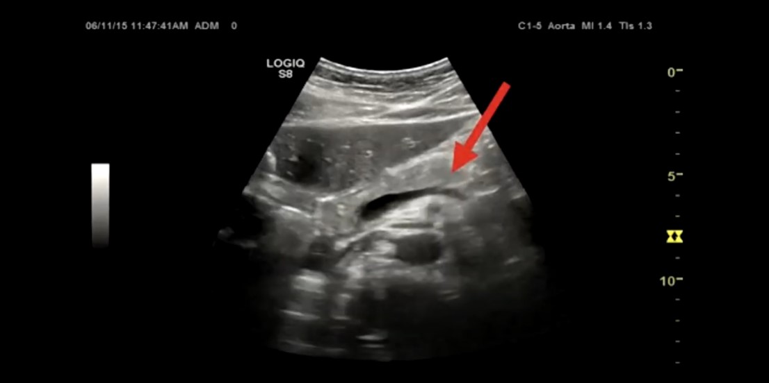


See all the echogenic thrombus.
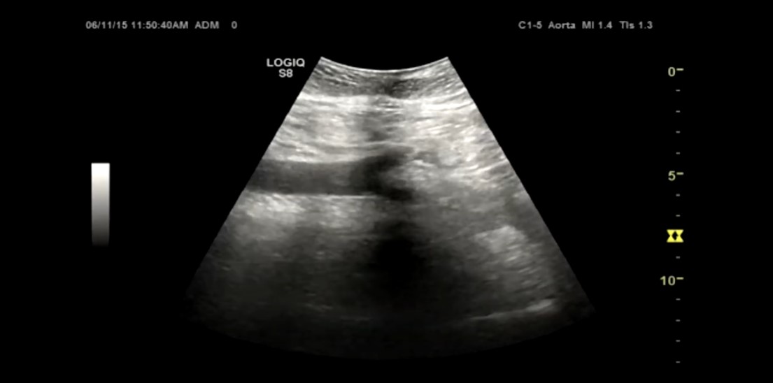
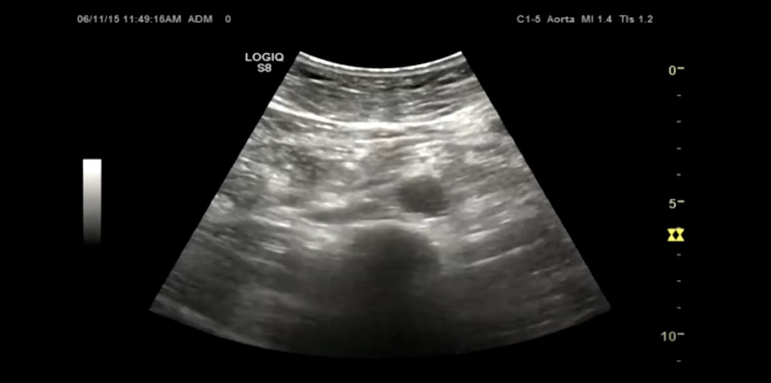
Another patient. Measured from outer wall to outer wall.
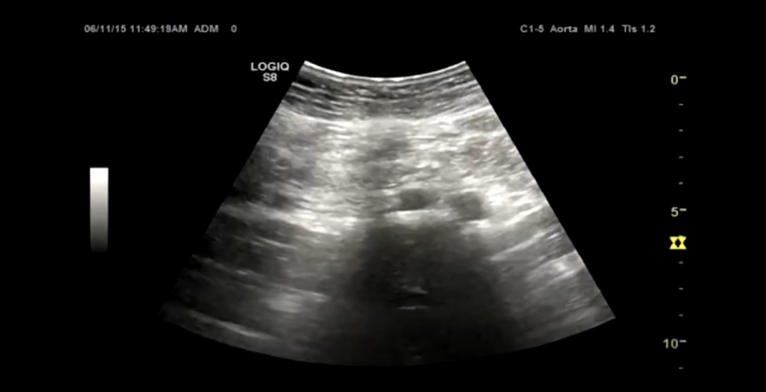
Another patient
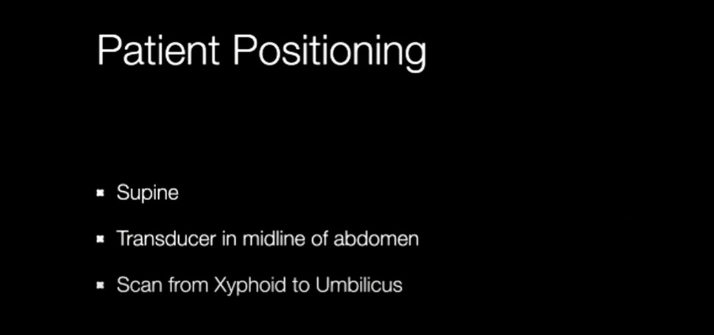
Scanning Protocol
Tranverse
Longitudinal
Transverse
Another tranverse view