First, be sure and review (and re-review) the excellent YouTube video, Ultrasound Lung Scanning Tutorial with Dr. Shane Arishenkoff – Clarius from Clarius Ultrasound. Only then, proceed to review the resources in today’s post.
In this post, I link to and embed MetroHealth Emergency Ultrasound‘s outstanding YouTube video by Dr. Greg Stoner, Pulmonary Ultrasound [The Blue Proocol], 27:03, May 25, 2022.
Although I will be placing slides from Dr. Stoner’s video tutorial in this post, I do this only to enhance my learning and to enable me to rapidly review the subject matter. [This blog is my study notes.]
I recommend that all readers use Dr. Stoner’s YouTube video to watch the video. By following his lecture on YouTube, you can follow the excellent YouTube (usually very accurate) autogenerated transcript.
In this lecture from our ultrasound lecture series Dr. Greg Stoner, DO (Emergency Ultrasound Fellow) talks about using bedside ultrasound to assess the lungs and pulmonary complaints? Follow us on Online and on Social Media:
Website: https://www.metrohealth.org/pocus
Twitter: https://twitter.com/MH_EMultrasound
Instagram: https://www.instagram.com/MH_EMUltras…
This is the official account of the Case Western Reserve University & MetroHealth Medical Center Emergency Medicine Ultrasound Division. Videos are not medical advice or views/policy of these institutions.
All that follows is from the above resource.
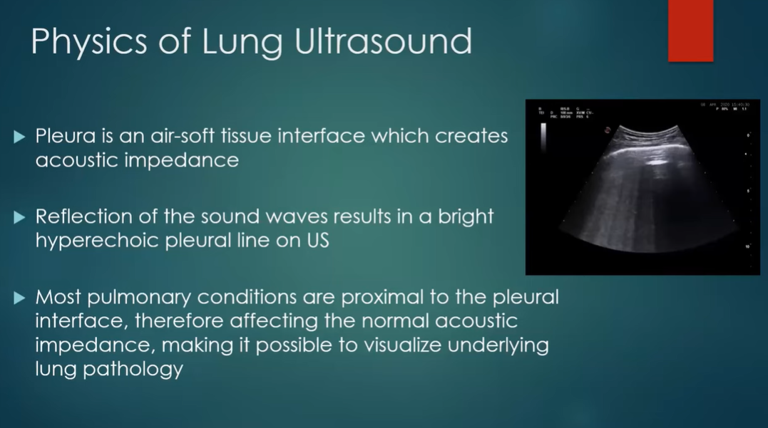
1:29
3:40
6:28
7:22
From the start of the lecture.
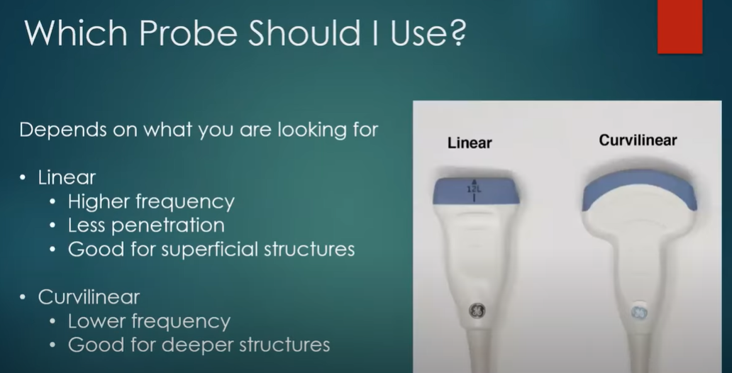
8:34
9:17
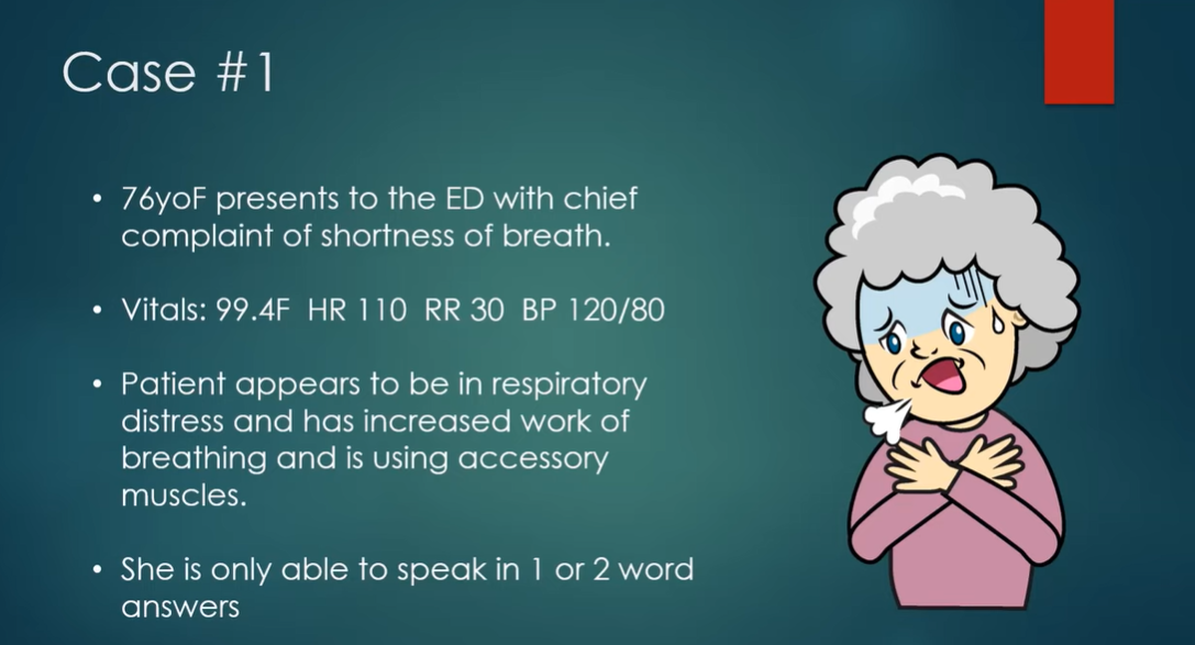
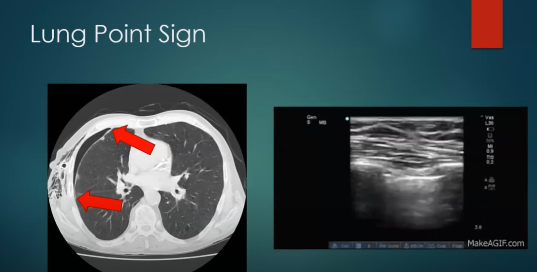
11:52
12:18
12:39
13:40
14:10-16:12 Below: Read the YouTube transcript.
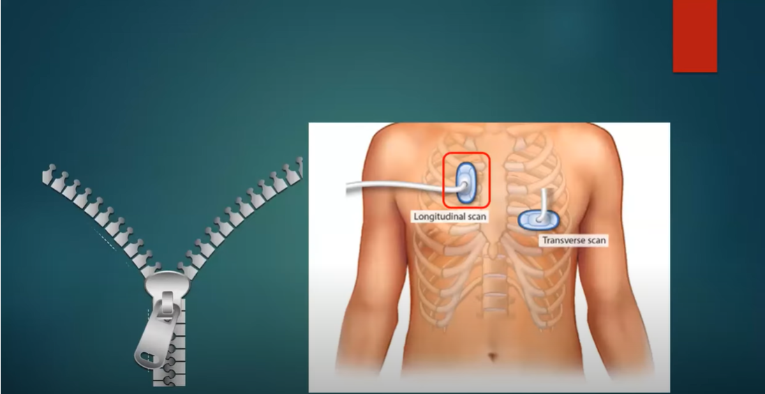
16:14 You are going to do a longitudinal scan.
17:52 Below: Read the YouTube transcript from 17:47 to 18:14.
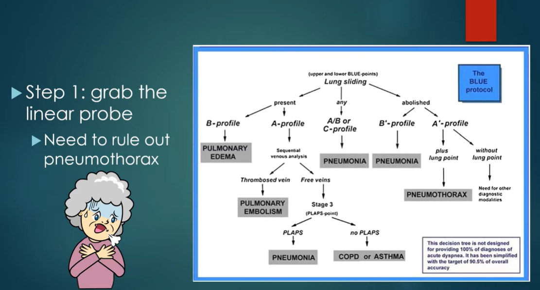

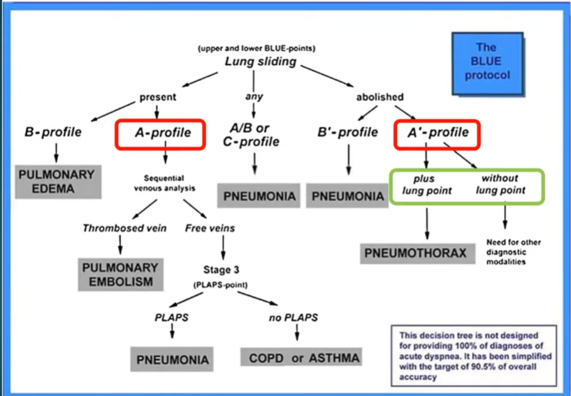
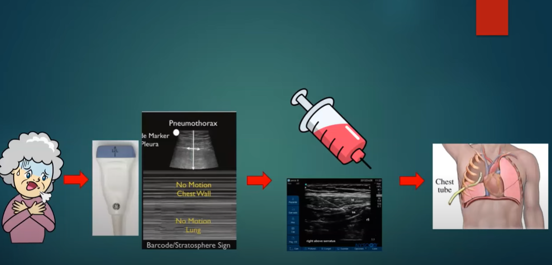
In summary for Case 1, following the BLUE protocol and your clinical information, you concluude that the patient’s seveere respiratory distress is caused by a pneumothorax. You treat it by providing good local anesthesia with a serratus anterior block* and place a chest tube.
*Please see
-
-
- Ultrasound Guided Serratus Anterior Block for Rib Fractures from MetroHealth Emrgency Ultrasound, Sept 22, 2021.
- Serratus Anterior Plane Block in the Emergency Department: A Case Series, Clin Pract Cases Emerg Med. 2020 Feb; 4(1): 21–25.
- Nerve Block Basics from MetroHealth Emergency Ultrasound, May 22, 2021.
-
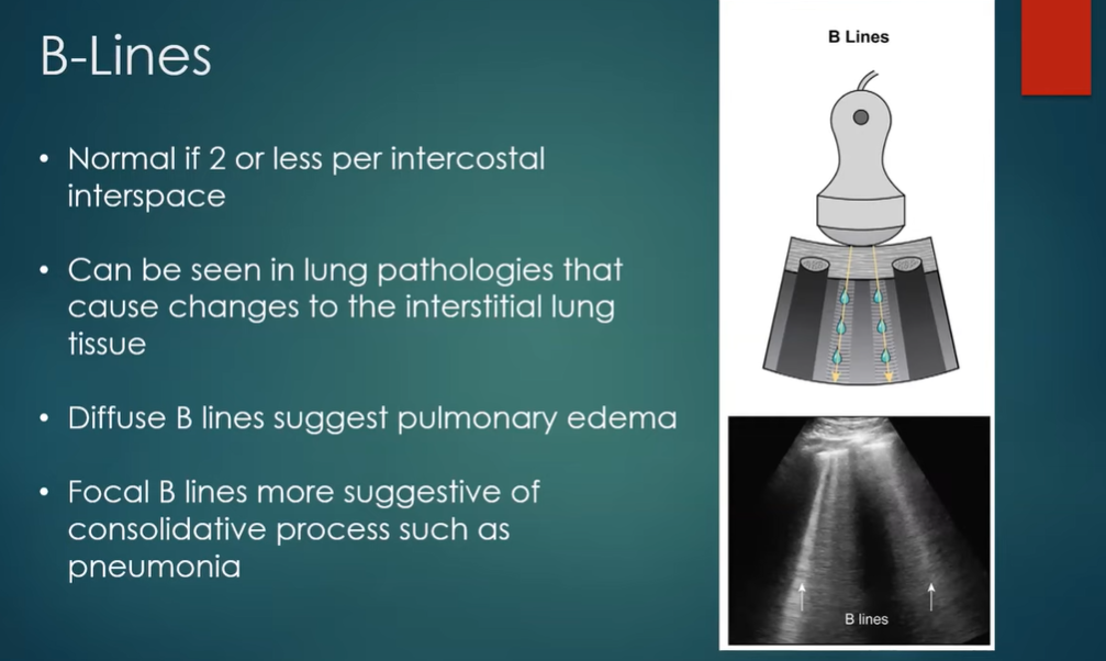
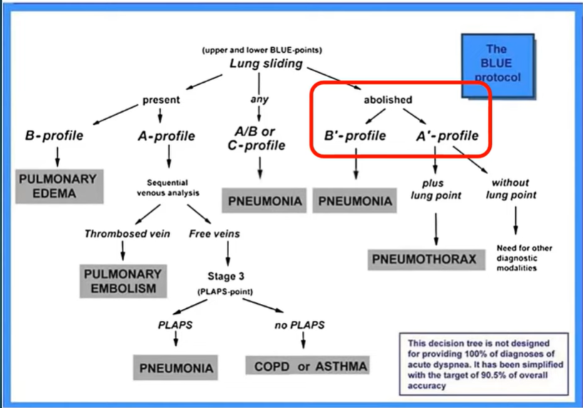
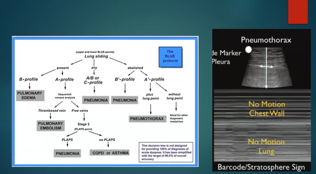
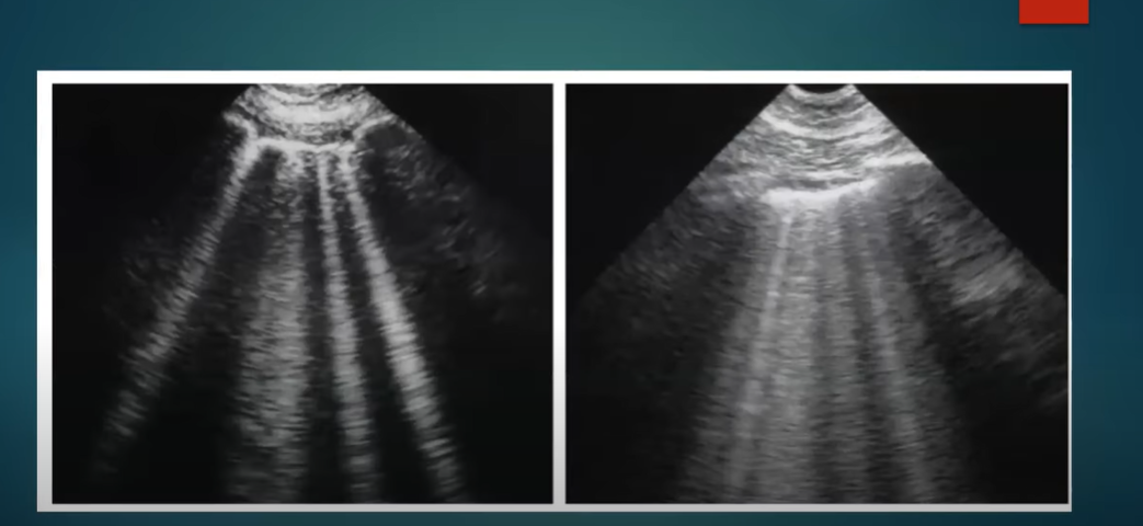
18:17:18:38 So, in this patient we see again no lung sliding motion [the barcode sign]. In this case, we see. . .18:56: . . . in this case we see B lines [the B’-profile] with no lung sliding and so this indicates pneumonia.

20:12
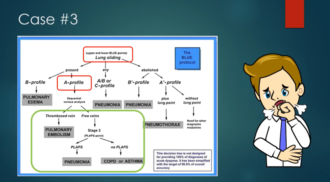
20:32 Case 3