Note: I originally posted this in May of 2015. I’ve moved it up to be with other recent RUSH posts.
Bedside Ultrasound in Resuscitation and the Rapid Ultrasound in
Shock Protocol [RUSH Exam] [Full Text PDF] Hindawi Publishing Corporation
Critical Care Research and Practice Volume 2012, Article ID 503254, 14 pages
This post contains a few quotes and the pictures from the above outstanding article–The BEST INTRODUCTION TO THE SUBJECT I’VE SEEN. And you will get a quick complete review of the exam just by looking at the pictures. But you need to read the whole article.
All that follows is from the above article:
“Early recognition and appropriate treatment of shock have been shown to decrease mortality [1, 2]. Incorporation of bedside ultrasound in patients with undifferentiated shock allows for rapid evaluation of reversible causes of shock and improves accurate diagnosis in undifferentiated hypotension [3]. Reflecting a trend to integrate ultrasound early into the care of the critically ill patient, multiple resuscitation protocols have been recently developed [4–26]. Each of these protocols combines many of the same core ultrasound elements, differing mainly in the priority of the exam sequence.
“In this paper, we will discuss two clinical scenarios of hypotension that will highlight how early integration of bedside ultrasound into clinical evaluation can assist in rapid and accurate diagnosis of shock [See article for the cases]. An easily learned and quickly performed shock ultrasound protocol, the RUSH exam (Rapid Ultrasound in Shock), will be applied in both
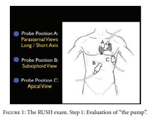
cases [19, 20]. The RUSH exam involves a 3-part bedside physiologic assessment simplified as “the pump,” “the tank,” and “the pipes.” Several other major resuscitation protocols will be compared to the RUSH exam to describe the core exam elements they share, as well as to demonstrate how they differ.”
Evaluate “the pump”:
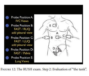
Evaluate “the tank”:
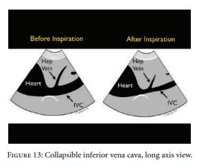
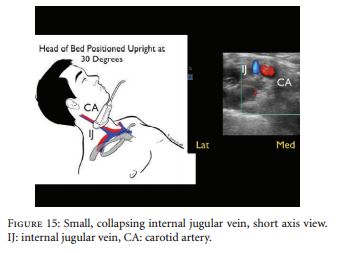
Inspiratory collapse:
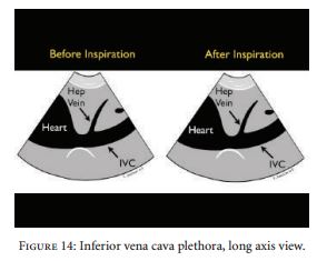
Plethora:
Evaluating the internal jugular vein:
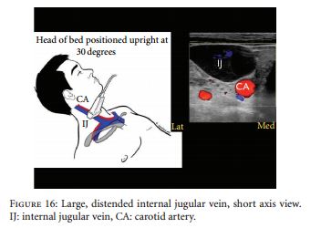
Large distended internal jugular vein:
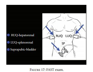
Evaluating “the tank”:
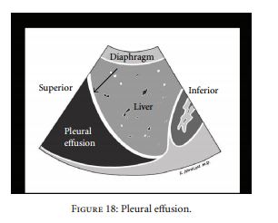
Pleural effusion:
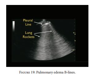
Pulmonary edema B-lines:
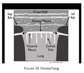
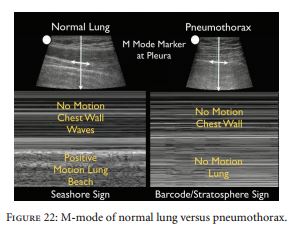
Look for lung sliding:
With pneumothorax there is no lung sliding:
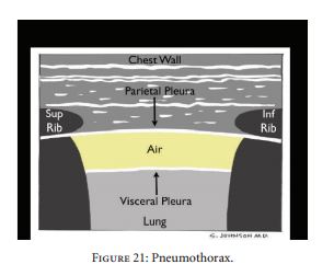
Normal lung with lung sliding vs. pneumothorax with no lung sliding:
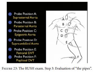
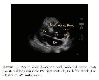
Evaluating “the pipes” (note that Probe position A evaluates the aortic arch and probe position B evaluates the aortic root):
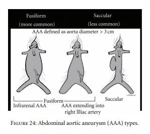
Types of abdominal aortic aneurysm:
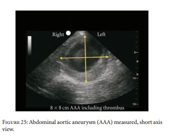
Abdominal aortic aneurysm:
Aortic root dilatation on parasternal long axis (left atrium is mislabeled RA)
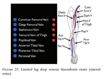
Exam for DVT:
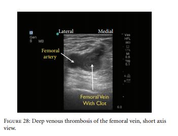
DVT of vemoral vein (vein non-compressible):
RUSH Protocol
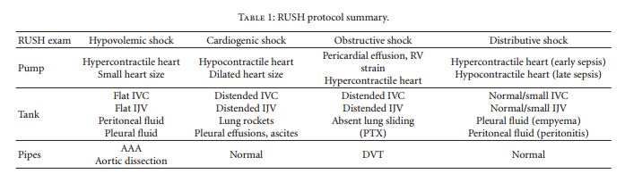
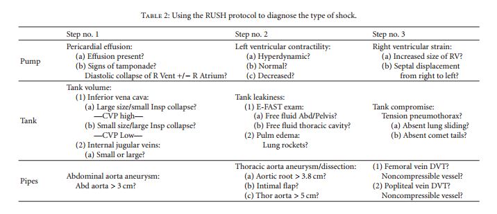
Using the RUSH protocol to diagnose the type of shock:
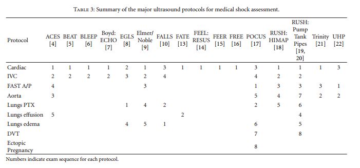
The different ultrasound protocols for the assessment of medical shock: