
The following video, Ultrasound Imaging of Hernia posted by 123radiology posted in 2015, is approx 43 minutes long but worth it, I think.
The slides of Part 1 are from 0 to 8:50, and cover the pathology of abdominal hernias.
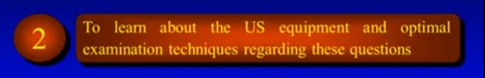
The slides of Part 2, are from 8:50 to 22:30, and cover ultrasound exam techniques.
The slides of Part 3, are from 22:30 to 27:24, and cover the ultrasound morphology of fixed and sliding hernias and asymptomatic and symptomatic hernias.
The slides of Part 4, are from 27:24 to , and cover bowel obstruction and vascular compression of the contents of hernias.
Section 2 runs from 8:50 to
Below are the four positions for abdominal hernia scanning:
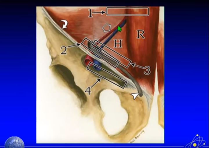
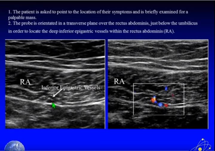
Below is the probe at position one:
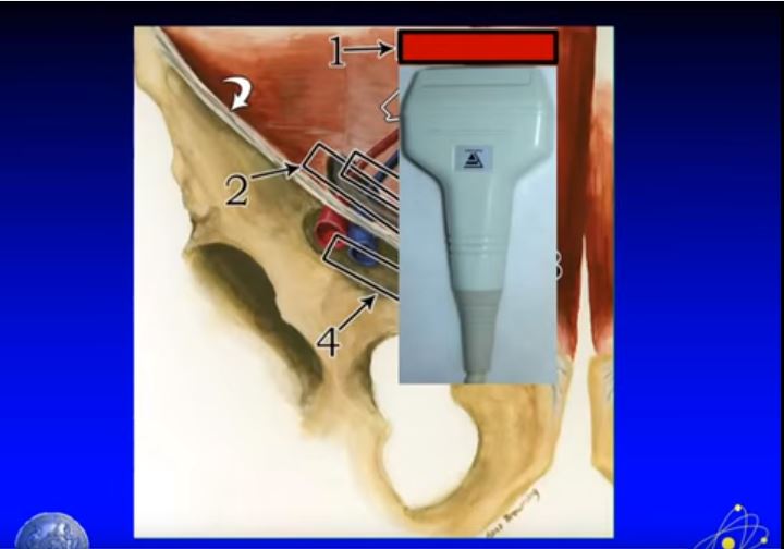
And below is what we see at position one:
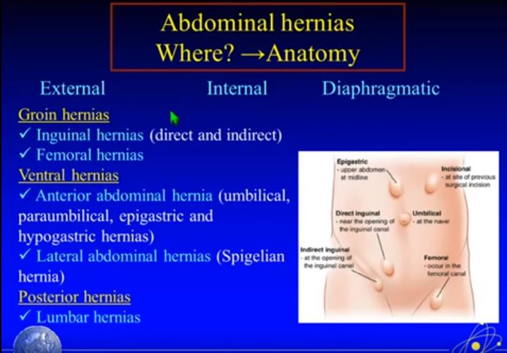
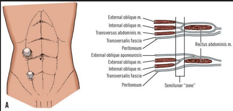
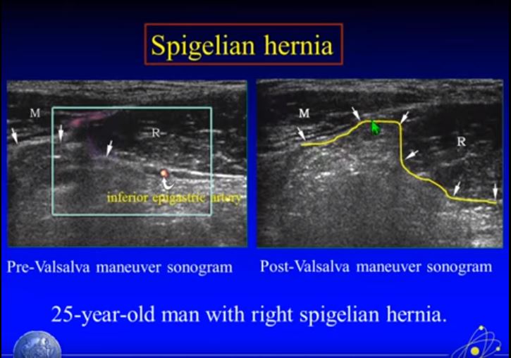
Position 1 is a good position for evaluating a Spigelian hernia* below: *”A Spigelian hernia (or lateral ventral hernia) is a hernia through the spigelian fascia, which is the aponeurotic layer between the rectus abdominis muscle medially, and the semilunar line laterally.”
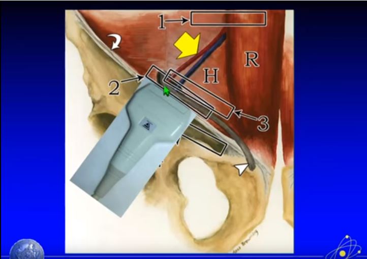
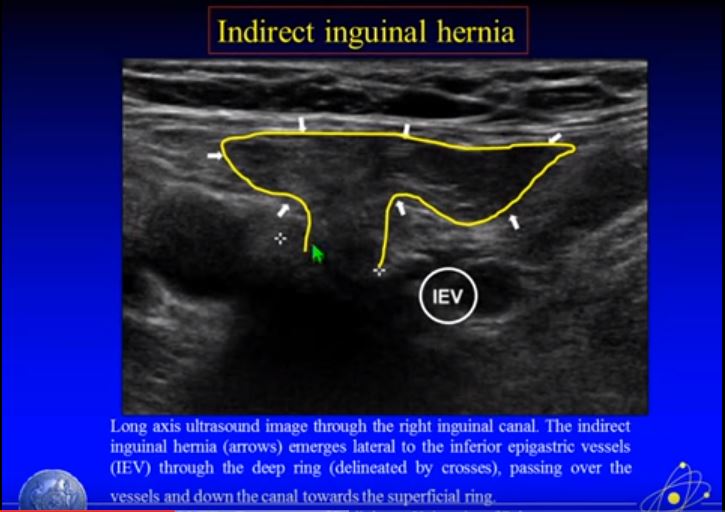
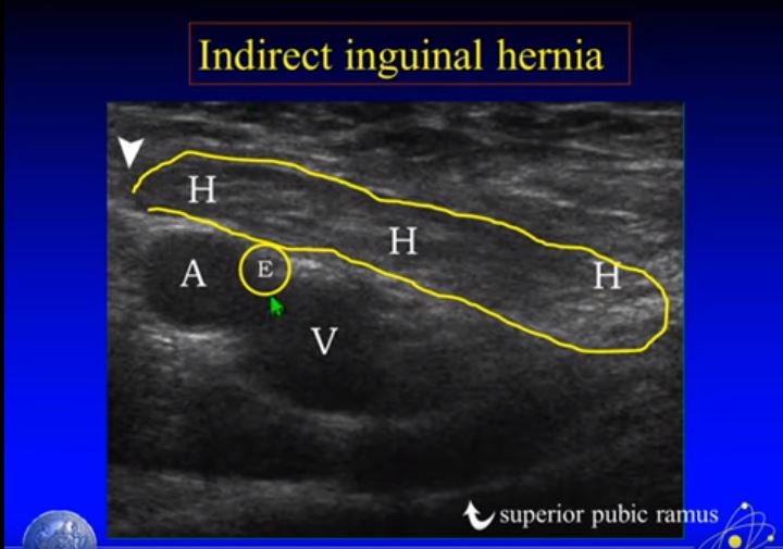
From Position 1 we move to position two by following the inferior epigastric vessels. And at position 2 we’re going to evaluate the inguinal canal:
here is some stuff
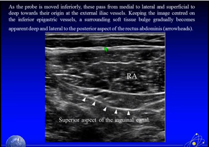
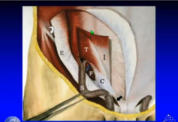
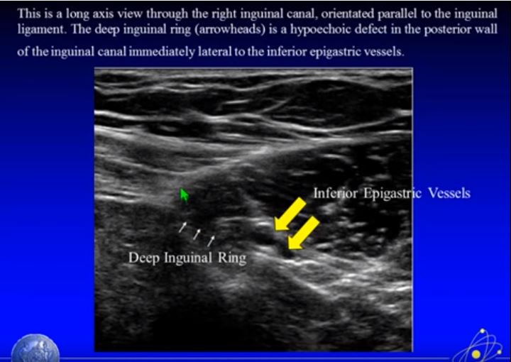
The Deep Inguinal Ring is lateral to Inferior Epigastric Vessels:
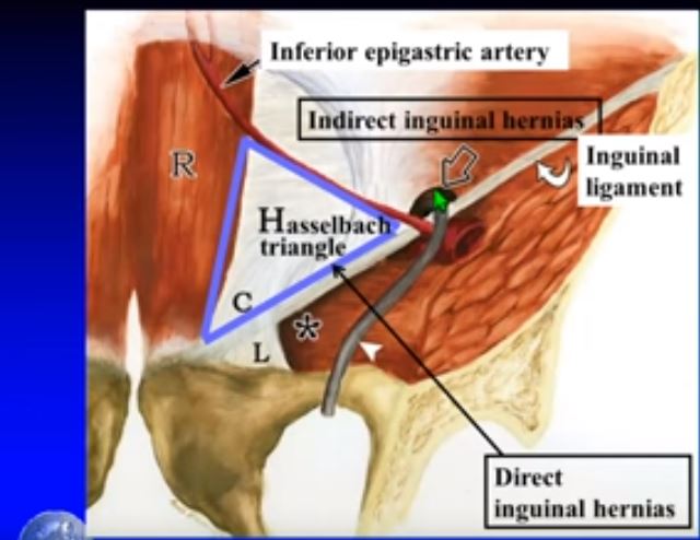
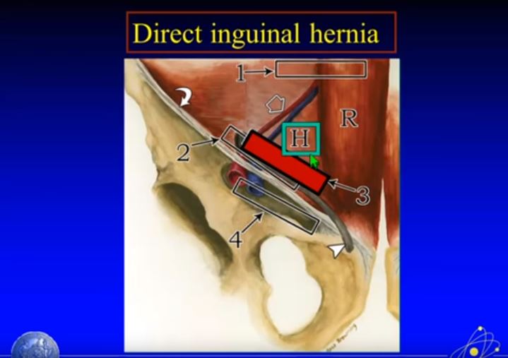
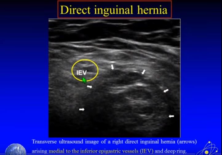
If we move a little bit medial to position 3, we are looking at the area of the Hasselbach triangle. And this is where we look for direct inguinal hernia.
Start here at 14:12
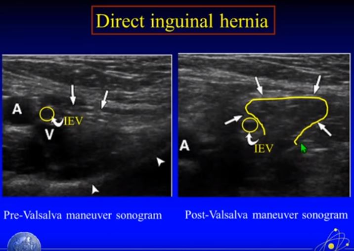
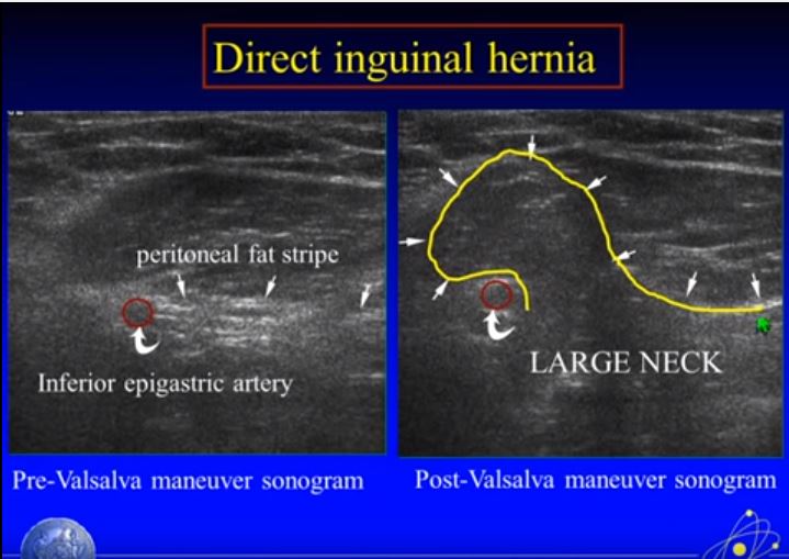
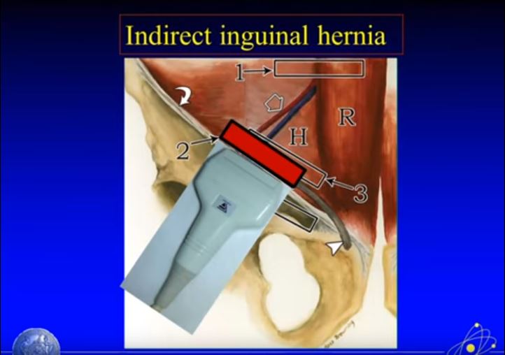
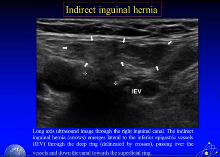
Below is a schematic of the inguinal region. Fig a shows the deep inguinal ring lateral to inferior epigastric vessels. Fig b below shows a direct inguinal hernia medial to the inferior epigastric vessels. wFig c shows an indirect inguinal hernia arising from the deep inguinal ring medial to the inferior epigastric vessels.
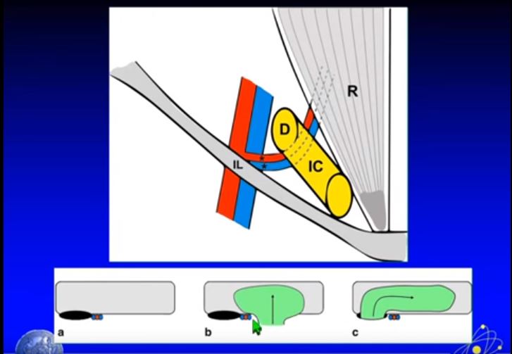
The direct hernia projects through the inguinal triangle. The indirect inguinal hernia projects through the internal inguinal ring.
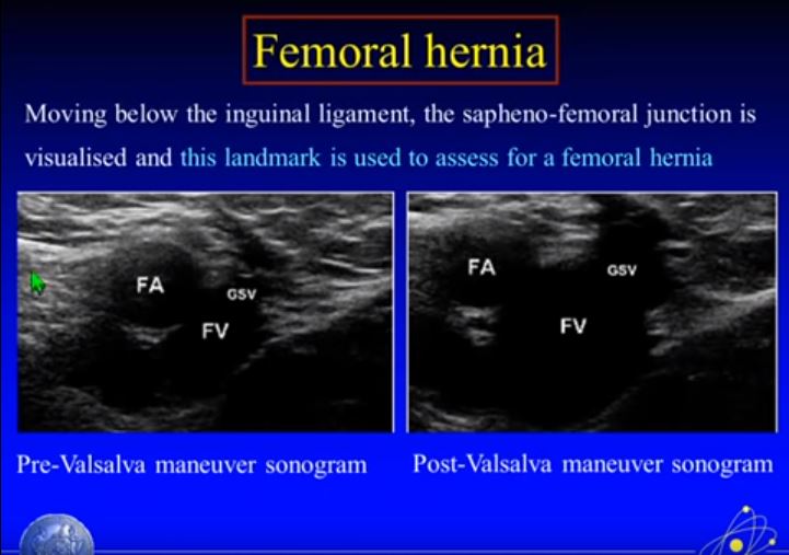
We now move to area 4 to evaluate for femoral hernia below. And we have to move our probe inferiorly to the inguinal ligament:
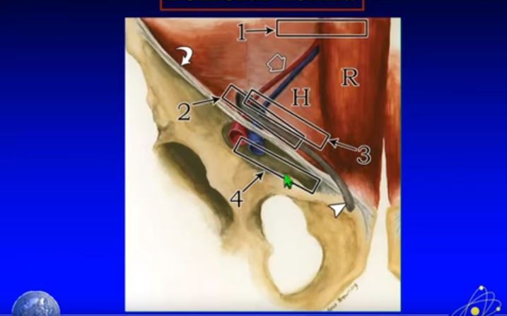
Again, we need to move the probe inferior to the inguinal ligament:
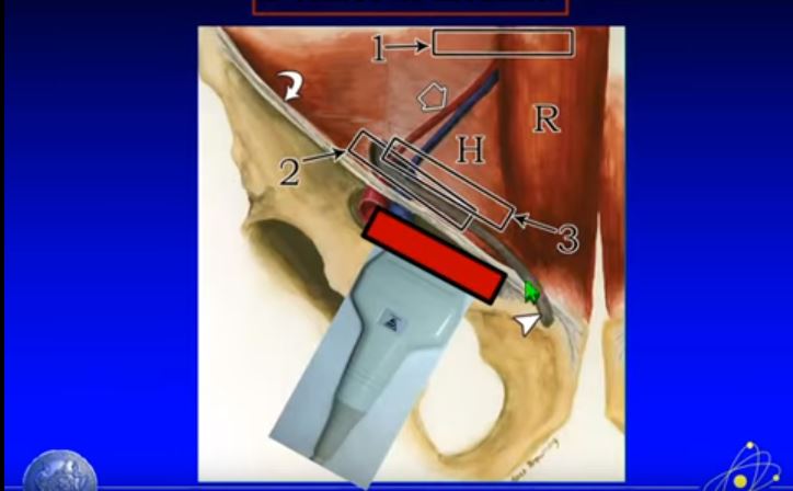
Here is the oblique view below:
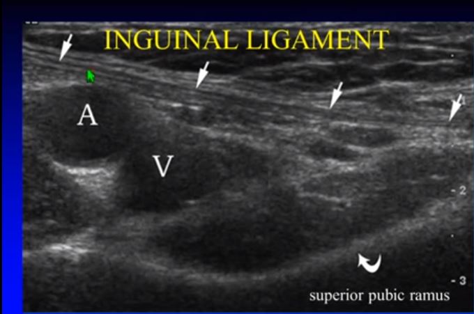
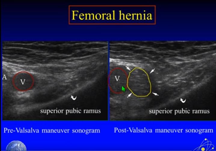
The femoral hernia lies medial to the femoral vein and below the inguinal ligament below:


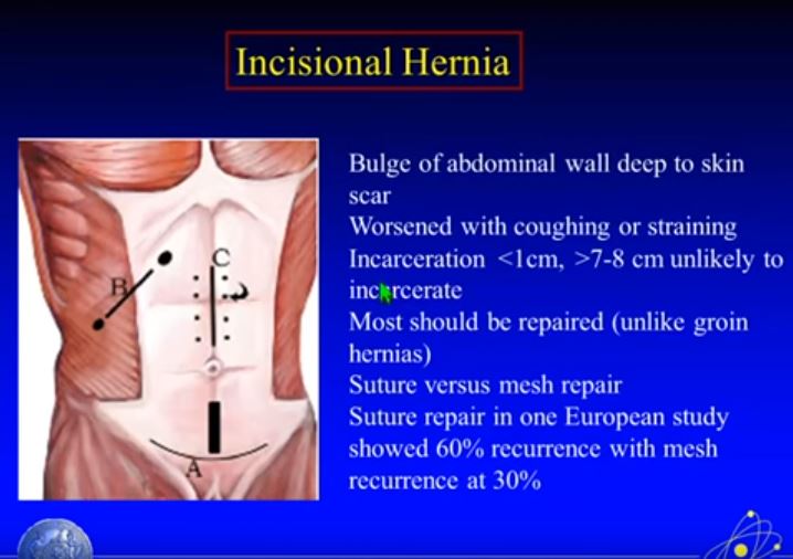
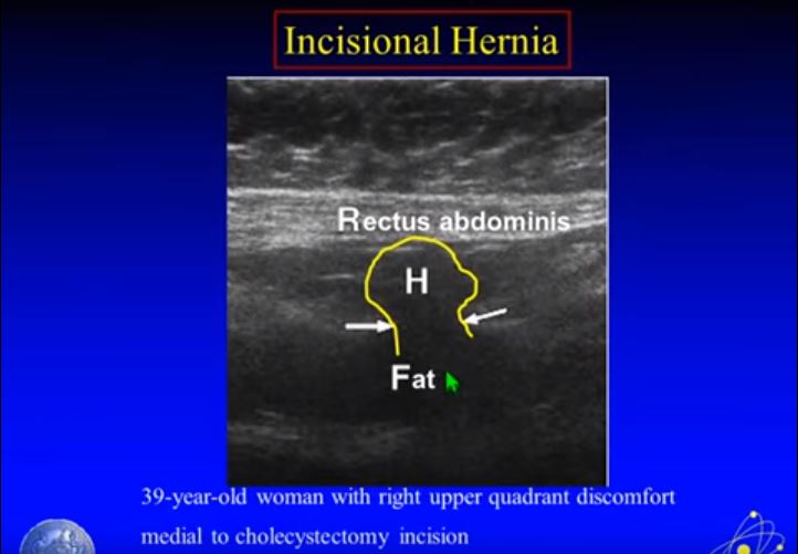
Finally, we have an incisional hernia:
48
49


