In this post, I link to and excerpt from The Curbsiders‘ #37: Out of Rhythm: SVT with Dr. Fahey. OCTOBER 27, 2021 By DR SAM MASUR
All that follows is from the above resource.
Summary
Does learning cardiology give you palpitations? Not to worry, Dr. Mike Fahey returns to address all your concerns about supraventricular tachycardia. He teaches us about cardiac conduction, Wolf-Parkinson-White, and more in this shocking episode.
SVT Pearls
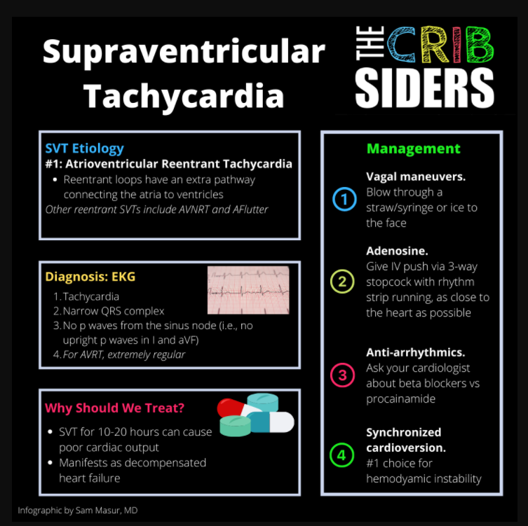
- The most common form of SVT in kids is Atrio-Ventricular Reentrant Tachycardia (AVRT), where there is an extra pathway that connects the atria and ventricles other than the AV node and His-Purkinje system.
- The most common EKG findings in retrograde AVRT (where the signal travels from ventricles to atria) are: narrow complex QRS, extremely regular QRS, and no upright P waves in leads I or aVF.
- The most effective vagal maneuver for children is often blowing into a partially occluded straw, which is a way to create a valsalva maneuver. For infants, Dr. Fahey recommends “ice to the face.”
- Adenosine will break every reentrant rhythm that involves the AV node.
- Adenosine should always be given with a rhythm strip running.
- The diagnosis of Wolff-Parkinson-White (WPW) is made purely with EKG and only needs to be captured once.
- Although rare, the risk of sudden cardiac death in WPW is due to an extremely fast electrical signal in atrial fibrillation. If the accessory pathway can conduct at high speeds, this atrial signal can cross to the ventricles, causing ventricular fibrillation.
- Do not give adenosine in patients with WPW (anterograde conduction across an accessory pathway) and atrial fibrillation.
- When children weigh at least 25kg, an electrophysiologist can ablate the accessory pathway in WPW.
SVT Notes
Cardiac Conduction System
Normal Sinus Rhythm
Conduction begins from the sinus node in the high right atrium. The sinus node has the fastest automaticity and it doesn’t need any stimulus to depolarize. After depolarization, the electrical stimulus travels through the atria towards the ventricles, but cannot pass across due to the fibrous ring electrically insulating the structures. The AV Node is the crossing point, where the stimulus waits a split second, and then rapidly travels down the His-Purkinje system to depolarize the ventricles in a very short amount of time. The signal would eventually make its way back to the atria, but it cannot due to the fibrous ring. Once depolarized, every myocyte goes into its refractory state, and the process starts all over again. If you work someone to their limits, the fastest physiologic heart rate is considered to be 220 – age (years).
If you see a narrow QRS, you know the electrical stimulus came through the AV node and traveled down a functional His-Purkinje system.
SVT Etiology
The most common form of SVT in kids is Atrio-Ventricular Reentrant Tachycardia (AVRT). In AVRT, there is an extra pathway (accessory) that connects the atria and ventricles other than the AV node and His-Purkinje system. Thus, electricity continues to loop through the atria and ventricles along the His-Purkinje system and accessory pathway. The prototypical cause of AVRT is Wolff-Parkinson-White Syndrome (WPW).
Another form of SVT that often gets confused with AVRT is Atrio-Ventricular Nodal Reentrant Tachycardia (AVNRT). In AVNRT, the reentrant loop happens within the AV node itself rather than through an extra pathway.
One of the rarest forms of reentrant SVT in pediatrics is Atrial Flutter, which is occasionally seen in infants. This reentry loop exists entirely within the atria.
Although less prevalent than reentrant tachycardias, the most common form of automatic tachycardia is Ectopic Atrial Tachycardia (EAT). In EAT, there is an electrical signal coming from another part of the atria that beats faster than the sinus node.
EKG
Ultimately, the diagnosis is contingent upon the EKG. The following are the most common EKG findings in retrograde AVRT:
- Narrow QRS complex
- Extremely regular QRS complex
- There is no discernable P wave coming from the sinus node (i.e. upright in lead I and aVF)
Note: it is very difficult to discern AVRT from AVNRT on EKG. This is a job for an electrophysiologist.
Other Testing
The remainder of testing is often completed once the arrhythmia has been treated (see below). Echocardiogram is recommended, as patients with SVT have a higher incidence of congenital heart disease (JAMA 2009). Dr. Fahey recommends checking a TSH in patients with atrial fibrillation, atrial flutter, or rarer forms of pediatric SVT.
Treatment of SVT
Why Should We Treat?
SVT is often not immediately dangerous, unless at very high heart rates (above maximum physiologic heart rate). The concern is directly proportional to the heart rate. In general, Dr. Fahey becomes worried about decreased cardiac output when a child has been in SVT for 10-20 hours or more. This would manifest as signs and symptoms of decompensated heart failure as above. For those children who find themselves with decreased cardiac output for a prolonged period of time, they can develop residual ventricular dysfunction, called tachycardia-induced cardiomyopathy.
Vagal Maneuvers
The vagus nerve works to slow sinus node automaticity and AV nodal conduction. Once AV nodal conduction is slowed, re-entrant loops involving the AV node will hit a new refractory period, causing the electrical signal to stop. Once that circuit breaks, the next electrical impulse will come from the automatic sinus node, the area with the most automaticity. Vagal maneuvers are exercises that can be performed to stimulate the vagus nerve and increase vagal tone. The following are commonly used maneuvers:
- Blow as hard as possible through a partially occluded straw. This mimics a valsalva maneuver, and is often the most effective according to Dr. Fahey
- For infants, “ice to the face” is one of the most common maneuvers. Grab a bag of ice, pour water in to create a slurry, and then place the bag on the upper portion of face with gentle pressure
- For infants, stimulate a gag reflex
- For infants, rectal stimulation
- For older children able to follow commands, the modified valsalva maneuver (blow into a syringe and invert legs) can be used (REVERT Trial 2015).
Note: most older children with previous episodes of SVT know works the best for them
Adenosine
Adenosine works by shutting down the AV node completely. Any reentrant tachyarrhythmia that utilizes the AV node as part of the circuit should be broken with adenosine. The following must be performed when giving adenosine:
- Must be given push IV with a 3-way stopcock.
- Give as close as possible to the heart due to its extremely short half life.
- Run a rhythm strip or EKG while giving it to identify any changes once the adenosine has been pushed.
- Adenosine should not be given to high-risk patients with WPW in atrial fibrillation due to the risk of shunting the fibrillating signal directly down the accessory pathway (see below).
If adenosine does not work, it is unlikely that there is a reentrant loop that includes the AV node. This often happens in atrial flutter or automatic tachycardias, such as EAT or atrial fibrillation.
Dr. Fahey’s decision to give more adenosine is based on the rhythm the patient is in. This is why the concurrent rhythm strip is so important. He will likely try one extra dose of the full adenosine just in case there was an unlucky aberrant beat that put the patient right back into the tachyarrhythmia.
Other Medications
If adenosine is ineffective, the conduction system of the heart must be changed in a more systemic manner. This is often called chemical cardioversion. Dr. Fahey’s next antiarrhythmic is often a beta blocker. Other types of antiarrhythmic agents are often chosen by local cardiologist preference, as there is little data comparing these medicines head-to-head in children. Examples include procainamide, sotalol, or flecainide. All antiarrhythmics are also pro-arrhythmic, so the more powerful the antiarrhythmic, the higher potential for triggering other nastier arrhythmias.
Electrical Cardioversion
In general, any hemodynamically unstable child in a supraventricular tachycardia should be electrically cardioverted as soon as possible. Electrical cardioversion works by delivering an electrical signal to every myocyte, causing them all to depolarize at the same time. Thus, every myocyte enters a refractory period, and then the most automatic signal in the heart starts back up again – the sinus node.
Electricity, in contrast to adenosine, will work for every reentrant rhythm as it affects every myocyte in the heart. In the stable patient, electrical cardioversion is often reserved for patients who can’t get IV access, but this is very rare.
Wolff-Parkinson-White (WPW)
Definition
A type of AVRT where the accessory pathway (extra pathway connecting atria to ventricles) can conduct electricity from atria to ventricles and/or ventricles to atria. The ability to conduct from the atria to ventricles is what makes WPW both unique and dangerous. It is also how WPW gets its hallmark EKG findings:
- Short PR interval – instead of pausing at the AV node, some of the electrical signal quickly crosses to the ventricles across the pathway, shortening the PR interval
- Delta wave – instead of the entire electrical stimulus traveling through the His-Purkinje system, some of the signal crosses the accessory pathway and directly depolarizes ventricular myocytes, initiating an early sloped QRS that eventually meets up with the remaining signal traveling the His-Purkinje system.
The diagnosis of WPW is successfully made with purely the EKG findings (and it only needs to be captured once!) Importantly, these findings will only be seen when the patient is out of SVT, as most patients are in retrograde AVRT.
Risks
Star here.