
In this post I link to and excerpt from The Cribsiders‘ [Link is to the complete list of Cribsiders’ podcasts] January 6, 2021 post by Dr. Justin Berk, #16: Kawasaki Disease with Recrudescent Guest Dr. Tremoulet.
Notes to myself: Review this [my] post rather than the excellent Cribsiders’ Episode #16 because I have included in this post some extra resources from the 2017 AHA Guidelines on Kawasaki Disease.
Also I recommend reviewing my post, 2017 AHA Guidelines On Kawasaki Disease – A Link And Excerpts. Posted on January 8, 2018. The post is for me a very efficient review of the AHA guidelines and reinforces the lessons of Dr. Tremoulet’s lecture.
Here are some of the key takeaways for me from Dr. Tremoulet’s lecture:
First, she tells us that the signs of Kawasaki Syndrome can come an go. And if a patient is seen over days by different providers who do not have access to previous physicians’ notes, he or she may not realize that the patient has Kawasaki Disease. The signs occurred sequentially and so no provider alone could make the diagnosis.
The key to avoiding the above problem, Dr. Tremoulet reminds us, is to take a careful history of the patient. Ask the parent if the patient has had any of the Kawasaki signs that have now resolved. If the parent has taken pictures of the patient during the illness, ask to review the pictures.
Dr. Tremoulet also reminds us that you don’t have to wait for the fever to be present for five days before you make the diagnosis of Kawasaki’s Disease.
If on day three of a fever, the patient meets the clinical criteria of Kawasaki Disease, then you can make the diagnosis at that time.
It is important to know that Kawasaki Disease is a clinical diagnosis that anyone can make.
Once you make the diagnosis, the patient needs to be admitted to the hospital for treatment.
Patients with untreated Kawasaki Disease have a 25% incidence of coronary artery aneurysms. With proper treatment started within ten days of the onset of fever, the incidence can be reduced to 5%.
In this post I link to and excerpt from The Cribsiders‘ [Link is to the complete list of Cribsiders’ podcasts] January 6, 2021 post by Dr. Justin Berk, #16: Kawasaki Disease with Recrudescent Guest Dr. Tremoulet.
Here are excerpts:
DON’T BE RASH, MAKE THE DIAGNOSIS!
Summary
With a persistent fever, Kawasaki Disease must be on your differential, but have you not seen many cases or had difficulty with the diagnosis? Look no further, as we bring an expert to walk us through the diagnosis and management of KD! We discuss Kawasaki Disease with Dr. Adriana Tremoulet who is the assistant director of the Kawasaki Disease Research Center at the University of California, San Diego. She has led numerous Phase I through III trials regarding the management of KD, and she walks us through the diagnosis and management of this fascinating disease!
What follows is from the 2017 AHA Guidelines on Kawasaki Disease:

Returning now to excerpts from the show notes of Cribsiders #16 Kawasaki Disease:
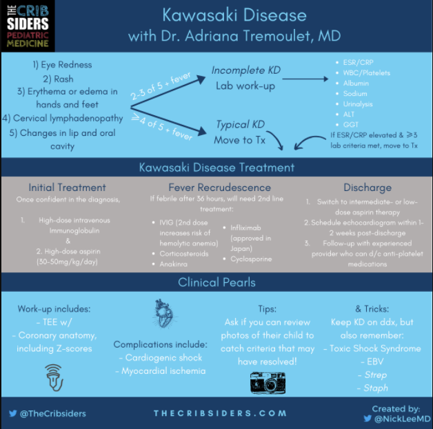
Core Criteria of Kawasaki Disease
The first step to diagnosing Kawasaki Disease (KD) is to include it on the differential that we are creating. Once you’ve done that, on your history and physical evaluate for the five core criteria (in addition to fever):
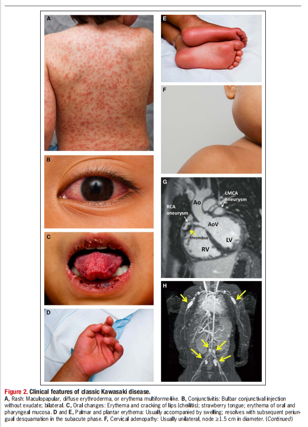
Eye redness
- Typically, both eyes become red at the same time without sick contacts or discharge
- “Limbic sparing” because it is technically injection and not conjunctivitis (no inflammation of conjunctiva in KD)
- Unilateral eye redness, one eye being more red than the other, sick contacts, or eye discharge is more suggestive of a acute viral etiology and not KD
Rash
- More prominent in the genitourinary area, so it is important to look underneath the diaper
- There is no classic KD rash, although it is not vesicular.
- Less likely KD (and more likely viral infection) if there are dots on the palms and soles
Erythema and edema in the hands and feet
- Can also present as a lack of unwillingness to walk in toddlers (sometimes due to concurrent hip arthritis)
- In undiagnosed KD, can eventually present as periungual peeling that starts underneath the nail bed
Cervical lymphadenopathy (≥1.5 cm in diameter; usually unilateral)
Changes in lips and oral cavity (eg strawberry tongue, cracked lips)
Less likely KD if there is:
- Palatal petechiae (Staph or Strep infection)
- Exudative pharyngitis (Staph or EBV infection)
Expert opinion: Dr. Tremoulet also recommends sitting down with the parents to review any photos that they may have of their child over the preceding days. This can help you identify criteria that may have come and gone prior to presentation!
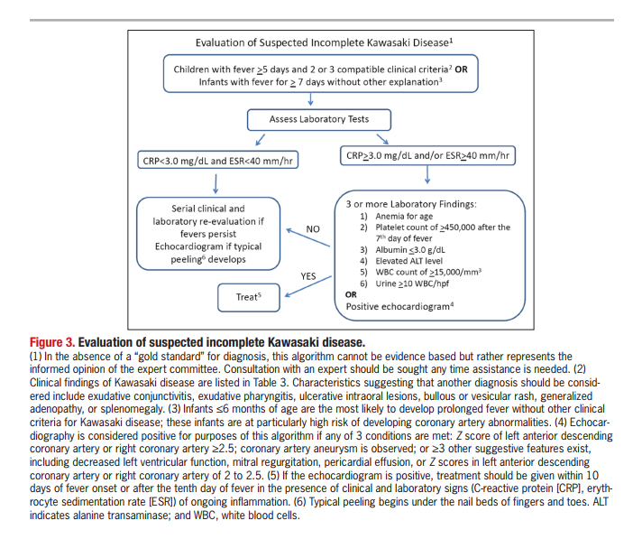
Incomplete Kawasaki Disease
When faced with incomplete Kawasaki Disease, the American Heart Association recommends further laboratory work-up to help characterize overall inflammation to aid in identifying children at risk for a coronary artery aneurysm.
- ESR/CRP
- WBC/Platelets
- Albumin
- Sodium
- Urinalysis
- ALT
- GGT (not in guidelines but expert recommendation)
What follows is from the 2017 AHA Guidelines on Kawasaki Disease [These are the values of the lab tests above]:
Now we are resuming excerpts from the show notes of Cribsiders #16 Kawasaki Disease:
Risk Factors for Kawasaki Disease
It is difficult to determine who is at greater risk for Kawasaki Disease since there is no mechanism or etiology fully known. Most of what is known is retrospectively established, but it includes the following groups are at a higher incidence of Kawasaki Disease or at higher risk for coronary aneurysms:
- Age < 6 months are at higher risk for CAAs (Salgado 2017)
- Extremes of anemia and platelet count (thrombocytopenia or thrombocytosis) can portend worse disease
- Earlier rise of inflammatory markers
- Self-identified race of patient and parents (in San Diego County; Tremoulet 2011)
Management of Kawasaki Disease
- Once confident in the diagnosis, give IVIG and initiate high-dose aspirin (30-50mg/kg/d divided q8 or q6)
- After 36 hours, monitor for recrudescence of fever
- If febrile after 36 hours, will need to give second-line therapy, which can include:
- IVIG (2nd dose increases risk of hemolytic anemia)
- Steroids
- Anakinra
- Infliximab
- Approved for Kawasaki Disease in Japan (but not in the US)
Upon Discharge
- Switch to intermediate- or low-dose aspirin after discharge
- If indicated, ensure that the patient is on the appropriate anticoagulation:
- If TTE Z-score* ≥ 5 and < 10, consider dual antiplatelet therapy (with aspirin and clopidogrel)
- If TTE Z-score* ≥ 10, therapeutic anticoagulation (e.g. enoxaparin) and low-dose aspirin
- Coordination of out-patient follow-up:
- Echocardiogram within one to two weeks post-discharge, which may require sedation
- Will need to determine when antiplatelet therapy can be discontinued
* For information about the meaning of Z-scores see The use of Z-scores in paediatric cardiology [PubMed Abstract] [Full Text HTML]. Ann Pediatr Cardiol. 2012 Jul-Dec; 5(2): 179–184.
COVID-19, MIS-C, & Kawasaki Disease
It can be difficult to tell the difference between the two, and it is possible that there may be overlap between the two syndromes. If you need a refresher on MIS-C, see Episode #5 with Dr. Tremoulet! MIS-C tends to have more frequent follow-up due to their severe myocardial dysfunction.


