
In this post I link to and embed three YouTube videos on using POCUS for Airway Examination.
Airway Ultrasound, Sept 11, 2017
This brief video [3:55] demonstrates an excellent way to use POCUS to locate the cricoid membrane for cricothyrotomy.
0:18 The probe is just distal to the sternal notch.
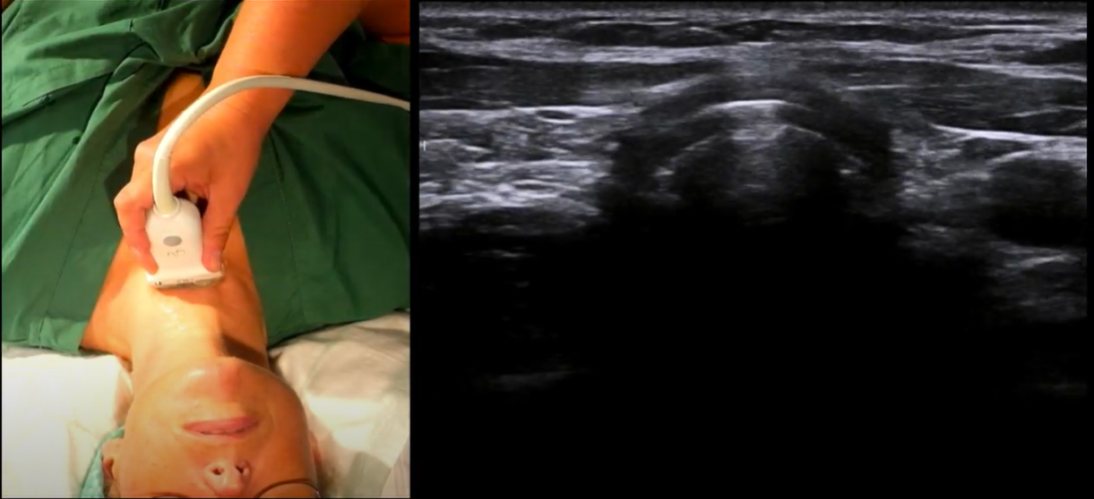
0:19 The trachea is here marked in orange. The trachea is air-filled which makes it hypoechoic and therefore dark.

0:30 The tracheal ring, which we here mark in red, is also hypoechoic.

0:38 As we move the probe cranially we see a series of tracheal rings.

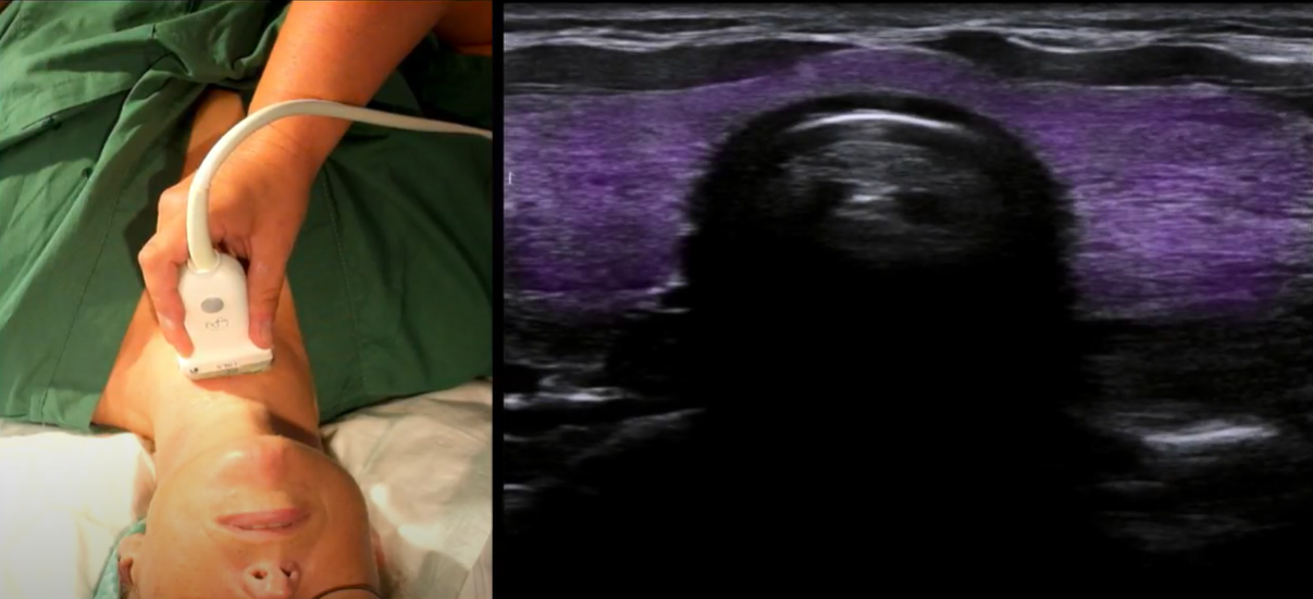
0:44 And [as we continue moving caudally] we now approach (encounter) our first anatomically important landmark, the thyroid gland.
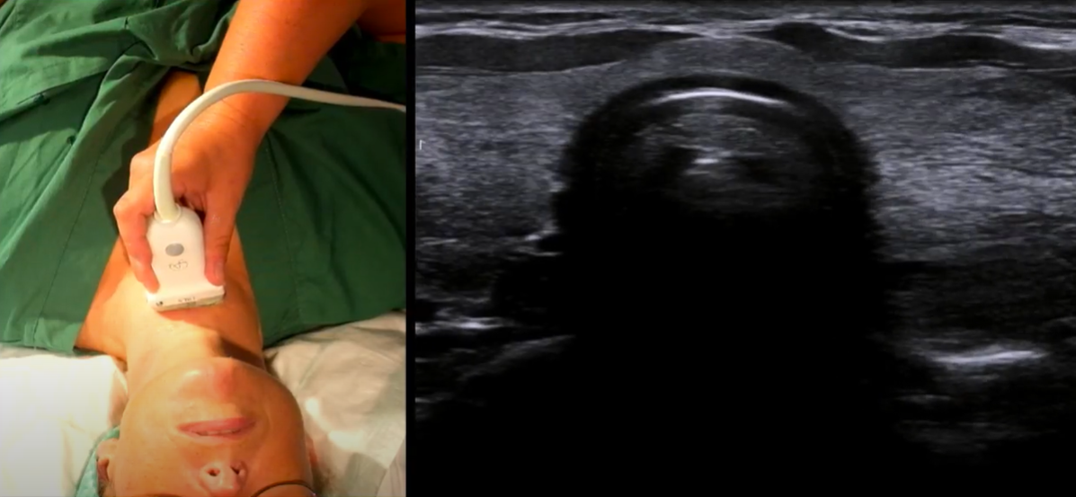
0:47 Here the thyroid gland is marked in purple.
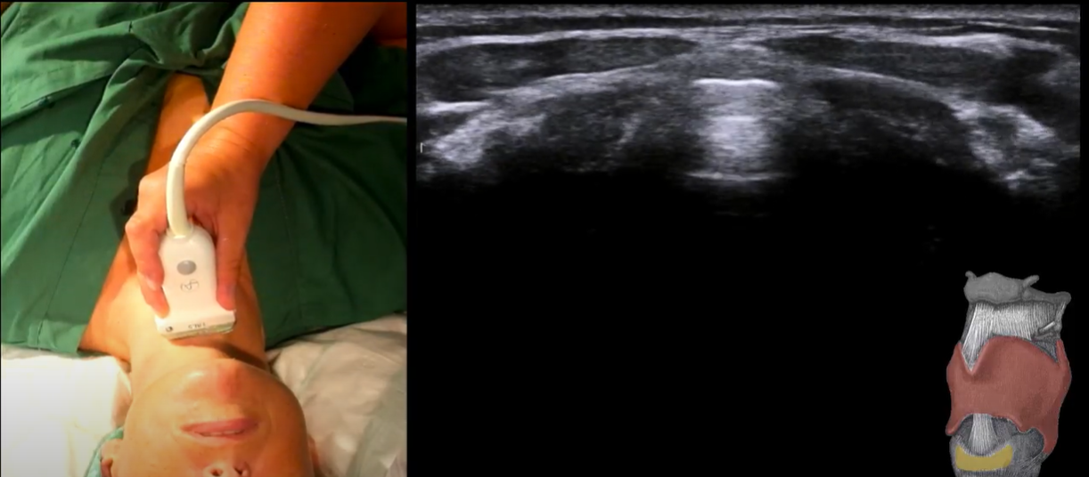
1:00 The next structure to appear is the cricoid cartilage.

1:03 Here the cricoid cartilage is highlighted in yellow. It is a horseshoe-shaped slightly larger ring.

1:05 Just cranially to the cricoid cartilage we see the cricothyroid membrane which appears as a sharp white line with parallel lines underneath, so-called reverberation artifacts which appear when there is a distinct tissue air interface.*
*Just like the A-line reverberation artifacts we see in a normal ultrasound lung scan.
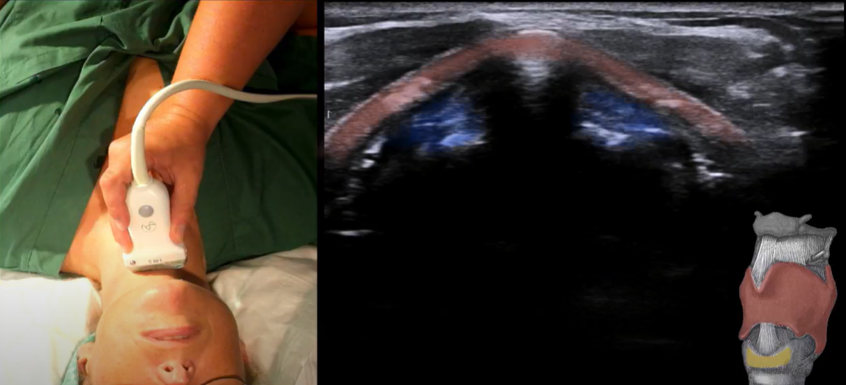
1:26 Further cranially we see the thyroid cartilage which appears as an upside down V.
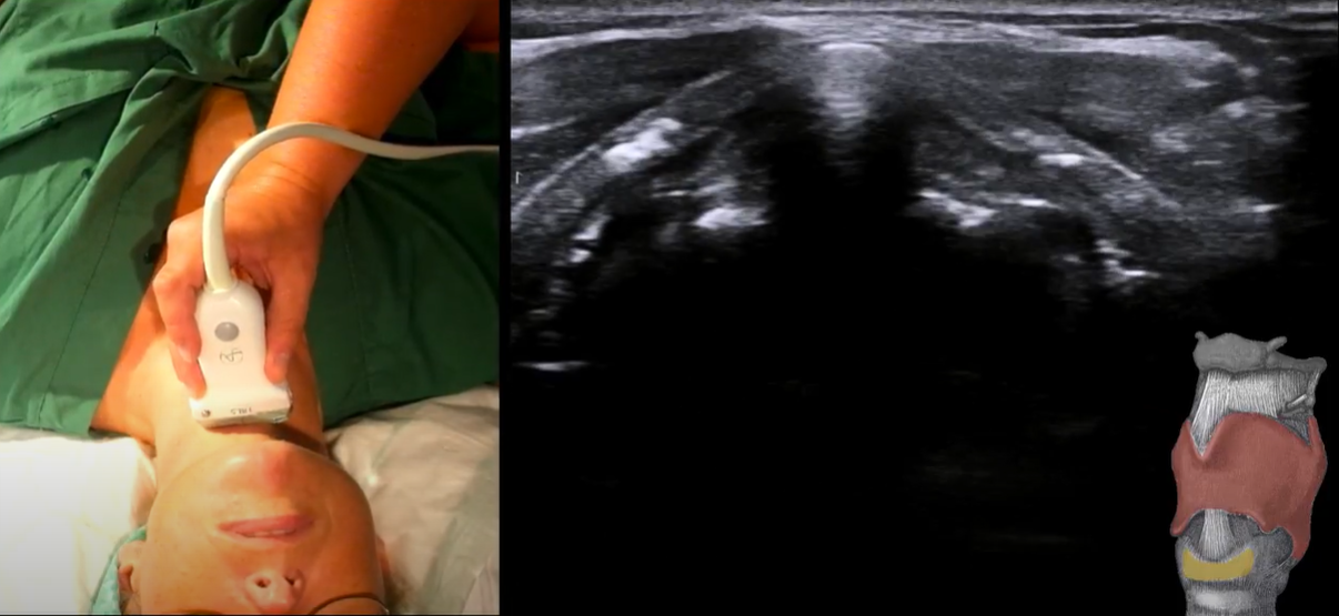
1:32 Here we have highlighted the thyroid cartilege in red. The muscles of the vocal cords are highlighted in blue in the image below.
1:55 As we move back caudally on the neck, we see the strucures again.
2:52 As we turn to the probe to a longitudinal direction, we see the tracheal rings as a string of black pearls.
fig14 when server ready.
3:02 As we move the probe crainially, we come to the cricoid cartilege.
fig15 when server ready
3:07 And here we see the cricoid cartilege colored in yellow with the thyroid cartilege and the cricothyroid membrane.
fig16 when server
3:20 – 3:45 Here we see how to determine the location of the cricothyroid membrane.
fig17 when server is available
fig18 when server is available.
Tutorial: how to perform an airway exam using POCUS, August 10, 2016.
The image below is a longitudinal view.


