
Earlier this year, I recertified in Advanced Cardiac Life Support. And the management of Torsade has always been somewhat unclear to me. But Dr. Farkas has three great resources on the subject and I will be using them and additional resources in this post, my study notes [As I remind occasional visitors this entire blog is my peripheral brain where I post my study notes and medical resources]:
- Torsades de Pointes Posted on November 22, 2016 from the Internet Book of Critical Care by Dr. Josh Farkas
- IBCC Episode 12 Podcast – Torsades De Pointes
- by Dr. Josh Farkas
- PulmCrit- A better approach to Torsade de Pointest July 2, 2018 by Dr. Josh Farkas
So I started my review of Torsades by listening to Dr. Farkas great podcast on the subject, IBCC Episode 12 Podcast – Torsades De Pointes. The podcast is approximately 16 minutes. It is a super review.
And then I reviewed the Internet Book of Critical Care post on Torsades De Pointes. All that follows is excerpted from the IBCC post:
getting some definitions straight
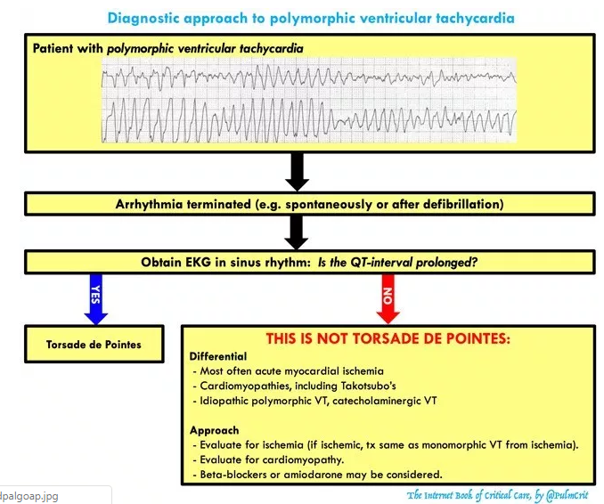
- Polymorphic ventricular tachycardia*
- Defined as ventricular tachycardia with varying QRS amplitude.
- This is commonly referred to as torsades de pointes, but it’s actually not the same thing. Polymorphic ventricular tachycardia may be caused by several etiologies (e.g. congenital QT prolongation, acquired QT prolongation, ischemia, Takotsubo’s cardiomyopathy).
- Torsades de Pointes*
- Torsades is defined as the combination of polymorphic ventricular tachycardia plus a prolonged QT-interval.
- Torsades can be caused by either congenital long-QT syndrome or acquired long-QT syndrome (due to electrolyte abnormalities and/or medications). The vast majority of torsades results from acquired long-QT syndrome, which is the focus of this chapter.
* For additional information on how to distinguish these two causes of polymorphic VTs see Resource (3) Polymorphic Ventricular Tachycardia Saturday October 12, 2013 from Dr. Smith’s ECG Blog
clinical presentations
- Depending on the length of the episode and whether torsades degenerates into ventricular fibrillation, clinical presentations include:
- Syncope or presyncope
- “Seizure” episode(s)
- Cardiac arrest
- Telemetry monitoring during an episode makes the diagnosis more straightforward.
EKG differential diagnosis: things that masquerade as torsades
- Severe hyperkalemia with “sine wave” pattern
- Coarse ventricular fibrillation
- Atrial fibrillation with Wolf Parkinson White (varying morphology may create polymorphic appearance)
- Polymorphic ventricular tachycardia with normal QT interval (may be caused by acute myocardial ischemia, Takotsubo cardiomyopathy, or such genetic conditions as catecholaminergic VT*).
* See Resource (4) below (4) The Athlete With Catecholaminergic Polymorphic Ventricular Tachycardia for good brief article on this topic.
Tx #1: breaking active polymorphic VT
unstable: immediate defibrillation
- Patients with unstable polymorphic VT need immediate defibrillation.
- The defibrillator will often be unable to lock onto any QRS complexes, so you generally need to perform an unsynchronized defibrillation.
- Technically the appropriate amount of energy is 200J biphasic. However, it might also be reasonable to use more energy particularly in a larger patient (with the rationale that unsuccessful defibrillation could be very dangerous, potentially pushing the patient into ventricular fibrillation).
“stable polymorphic VT”
- Polymorphic VT is never truly stable. This is a short-lived transitional state which usually flips back into sinus rhythm, or less commonly degenerates into ventricular fibrillation. Even if the patient looks OK, they’re not really stable: attach pads and don’t leave the room.
- Given the risk of degeneration into VF, urgent defibrillation is reasonable if the patient shows any hint of instability.
- Intravenous magnesium is a reasonable treatment, but a practitioner should be in the patient’s room with a defibrillator ready to shock the patient.
- Give 2 grams (8 mM) IV magnesium sulfate over 10-15 minutes, repeat if no response. If the patient remains stable and the rhythm persists, repeat an EKG and consider the possibility of an alternative diagnosis.
- Note that most treatments associated with torsades are used for the prevention of VT, not breaking an episode of VT. Thus treatments such as isoproterenol or pacing have no role here.
Tx #2: Basic tx to prevent recurrent torsades
If the EKG shows a prolonged QT-interval, the patient is diagnosed with torsades. If you simply break torsades but do nothing else, it is likely to recur. The following therapies will prevent recurrence of ventricular tachycardia in this situation:
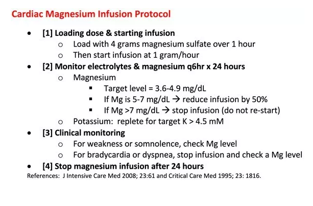
magnesium = 1st line therapy
- Patients with torsades should receive magnesium, even if they have a normal magnesium level.
- Four grams magnesium sulfate IV (16 mM) is a reasonable place to start. Unfortunately, if you stop after four grams then the magnesium level will fall over several hours and torsades may recur.4
- Additional magnesium should be provided based on the patient’s renal function:
- (a) If GFR > 30 ml/min, a magnesium infusion is useful (see protocol below).
- (b) If GFR < 30 ml/min, cycle magnesium levels and bolus intermittently to target a magnesium level of 3.5-5 mg/dL (1.5-2 mM).
- Magnesium has a very wide safety margin. A protocoled magnesium infusion may seem aggressive, but overall this is far safer than the risk of recurrent cardiac arrest.
- Below is a magnesium protocol (one version in American units, one in SI units).5,6 This will work best if pasted directly into the patient chart so that everyone is literally on the same page (physicians, nurses, and pharmacists).
- The rationale for a magnesium infusion is explored further here.
potassium
- Aggressively treat any hypokalemia, targeting a high-normal potassium level (>4.5 mEq/L).7
cessation of all QT-prolonging medications
- The medication list should be carefully reviewed for any medications which may prolong QT interval. Patients are often on several QT-prolonging medications, all of which should be stopped if possible.
- Evidence regarding which drugs cause torsades is extremely murky. In some cases it seems that drugs have been incorrectly maligned (e.g. azithromycin, olanzapine). One complicating factor is that there isn’t a simple relationship between QT prolongation and torsades (some drugs such as amiodarone increase the QT without causing much torsades).
- A comprehensive list of medications implicated in causing torsades is here.
[The rest of the IBCC post on Torsades has much more on the therapy of Torsades]
Additional Resources:
(1) The Table of Contents of The Internet Book Of Critical Care by Dr. Farkas.
(2) The Table Of Contents of PulmCrit by Dr. Farkas
(3) Polymorphic Ventricular Tachycardia Saturday October 12 from Dr. Smith’s ECG Blog
(3) Electrolyte Replacement Practice Management Guideline September 2016 from the Division of Trauma and Surgical Critical Care from the Vanderbuilt University Medical Center.
(4) The Athlete With Catecholaminergic Polymorphic Ventricular Tachycardia
Jul 28, 2017, Expert Analysis from the American College of Cardiology


