
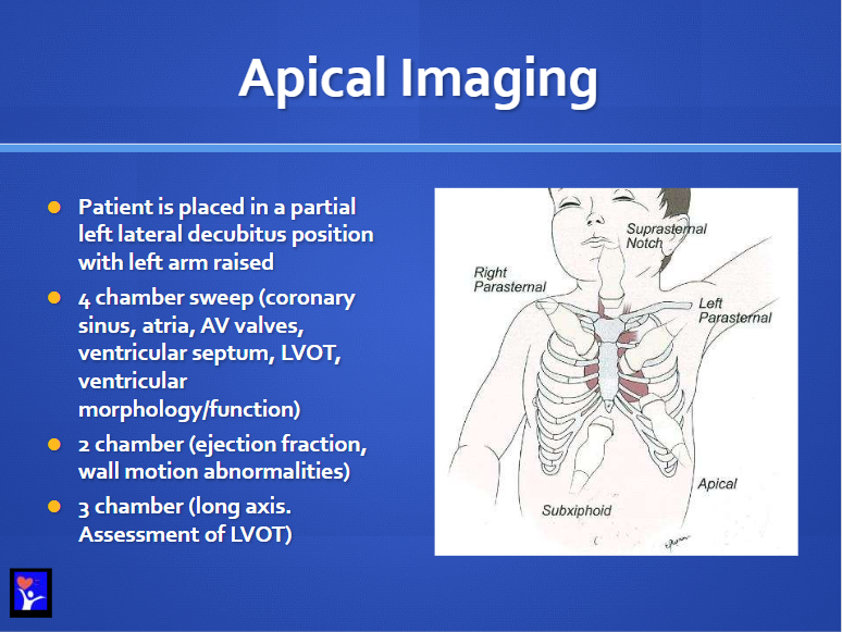
Apical Imaging: 31
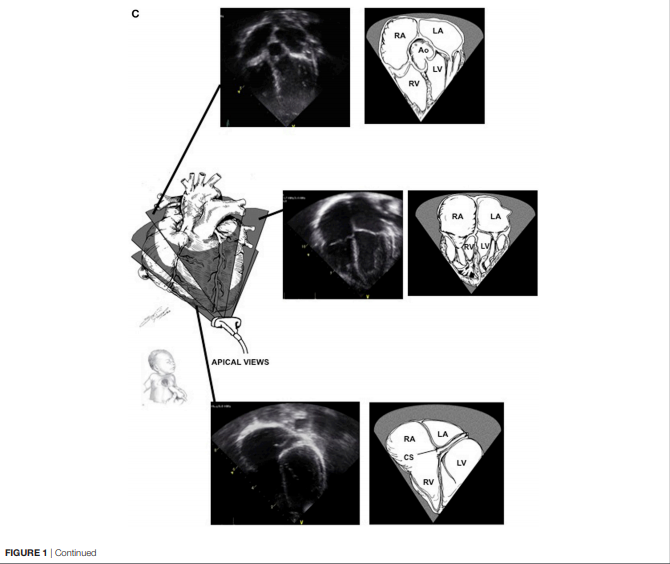
Apical Views [Fanning]: 32
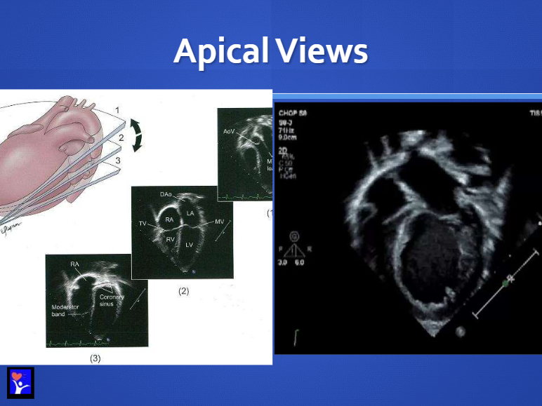
Apical Views from Basics of Functional echocardiography in Children and Neonates 2017:
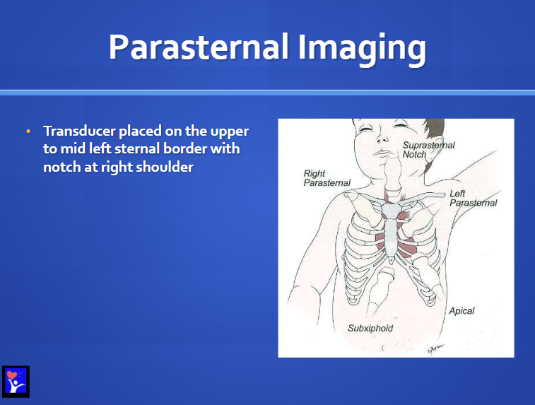
Parasternal Imaging: 37
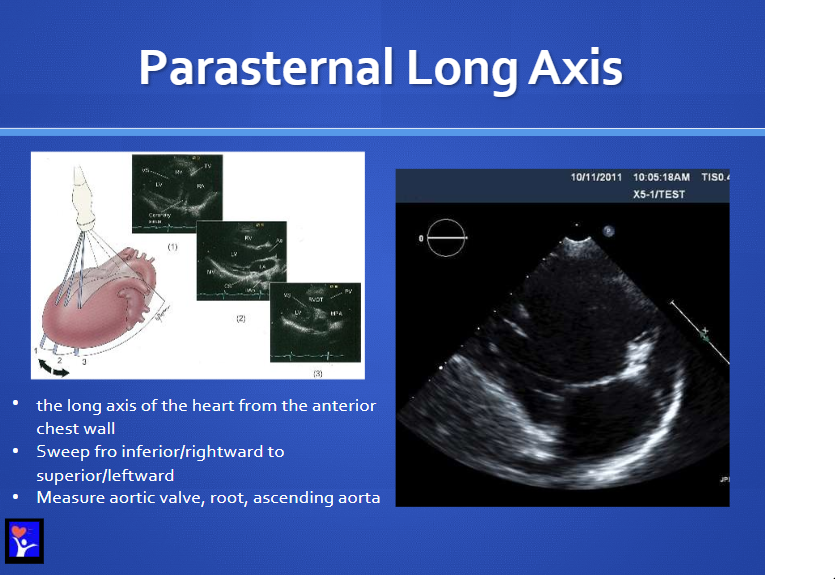
Parasternal Long Axis [Fanning]: 38
Transducer placed on the upper to mid left sternal border with the notch at the right shoulder. The long axis of the heart from the anterior chest wall
Sweep from inferior/rightward to superior/leftward
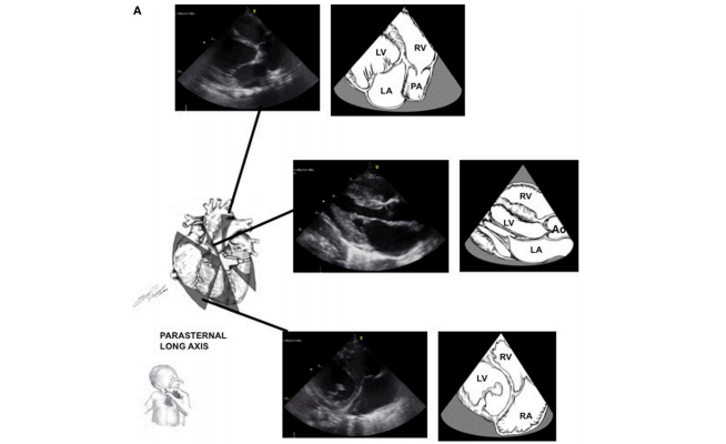
Parasternal Long Axis Views from Basics of Functional echocardiography in Children and Neonates 2017:
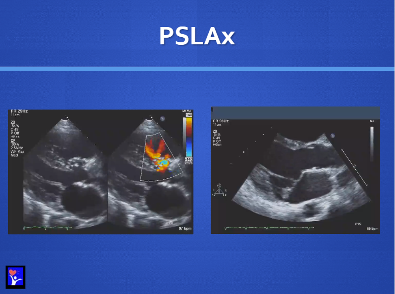
Parasternal Long Axis view showing a VSD in the area under the aortic valve in the membranous region: 39
Parasternal Short Axis [Fanning]: 40
[Transducer placed on the upper to mid left sternal border with the notch at the left shoulder.]
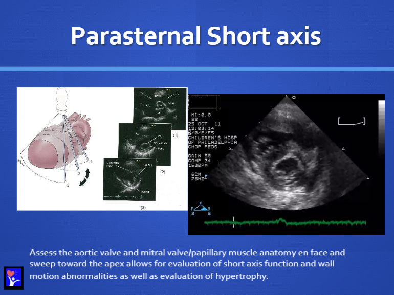
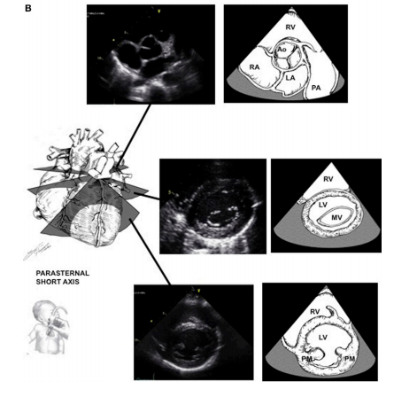
Parasternal Short Axis Fanning from Basics of Functional echocardiography in Children and Neonates 2017:
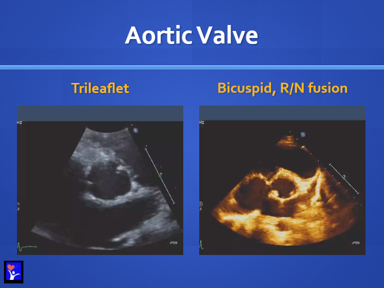
Aortic Valve Morphology (Parasternal Short Axis) : 41
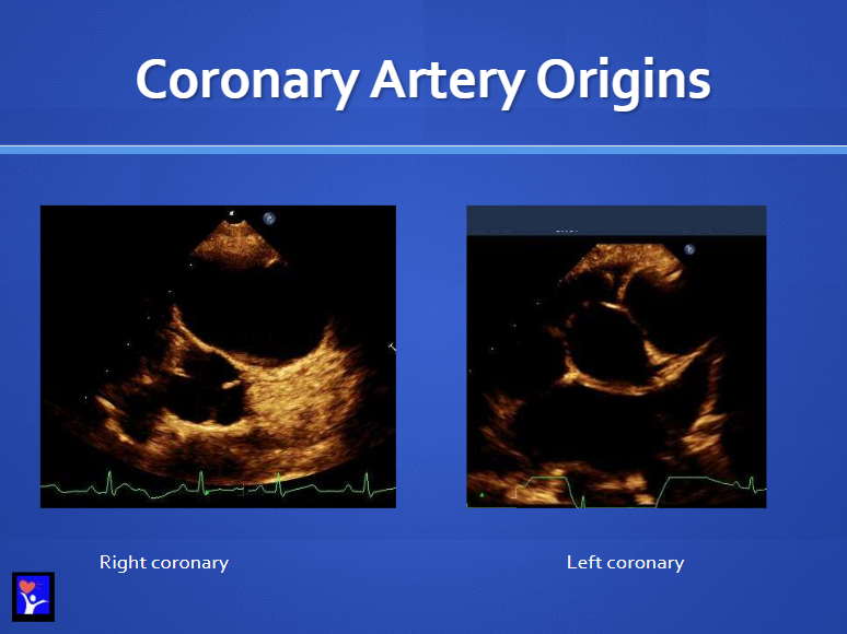
Coronary Artery Origins (Parasternal Long Axis): 42
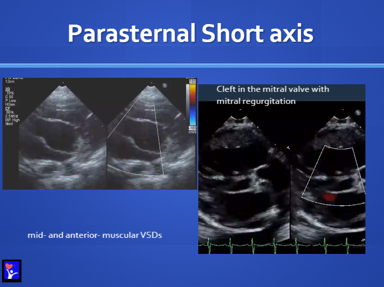
Parasternal Short Axis at the level of the mitral valve: 43
Pulmonary arteries view: 44
Position the probe slightly superior and coronal in the left chest
Pant leg view of the pulmonary arteries.
Ductal View: 45
High left parasternal window with notch at 12-1 o’clock
Great image of the PDA entering the aorta
Ductal View: 46
You can measure the full length of the PDA. You can measure the aortic end and the pulmonary end.
The pulmonary end and the aortic end can have quite different measurements
The duct tends to close from the pulmonary end to the aortic end
Ductal View: 47+48
Don’t really understand these images nor the lecturer’s points. Need to review with a knowledgeable examiner.
Suprasternal Views: 49


