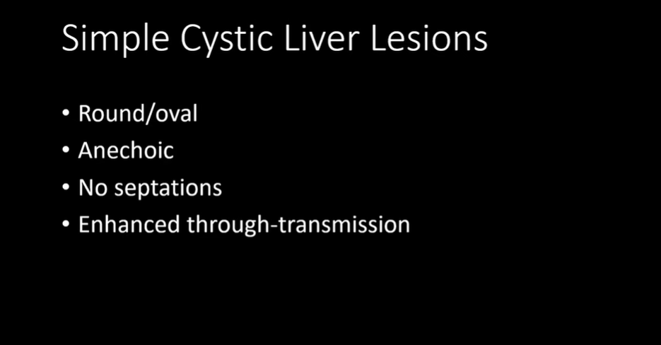
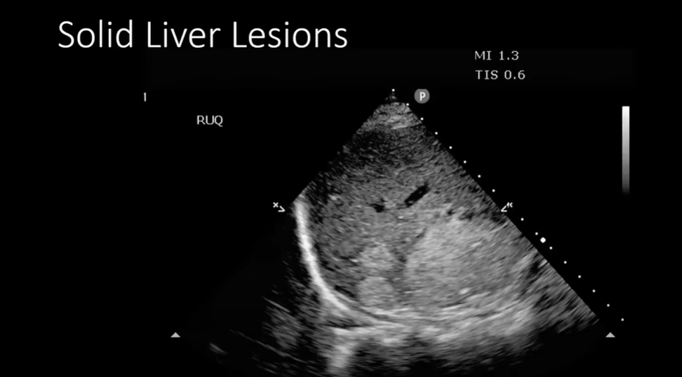
In this post, I review, link to, and embed the excellent YouTube lecture, Ultrasound of the Liver and Spleen from MetroHealth Emergency Ultrasound, 46:09, Oct 26, 2022, by Sandra Werner, MD.
Note to myself and my readers: The best way to quickly review this excellent teaching video is to watch the video on YouTube. By doing this, you can follow the video using the transcript. And as you go through the transcript you will be automatically reviewing the slides.
I put these resources on my medical education blog because the blog’s WordPress search function easily allows me to quickly find and access my study resources.
And copying and pasting the slides in a post (like this one) helps me reinforce my learning. But again, I recommend my readers review the video on YouTube so that they can follow along with the autogenerated (but usually very accurate) transcript.
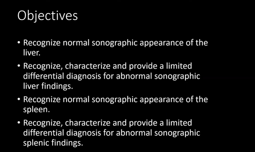
All that follows is from the above outstanding lecture by Sandra Werner, MD, Ultrasound of the Liver and Spleen.
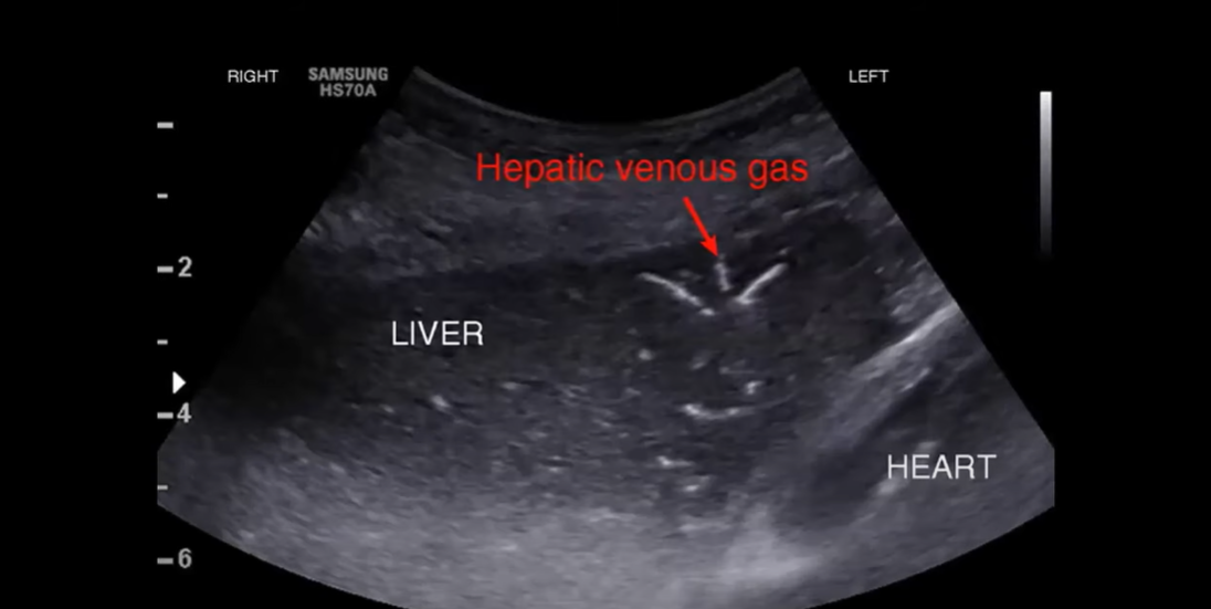
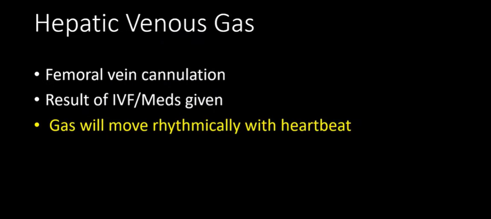
t
text
t
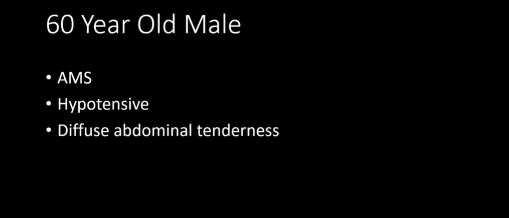
t
t
t
t
8:42
t
t
t
t
t
t
13:08:
13:45:
13:58 2nd case
14:13:
14:43
15:21
16:03
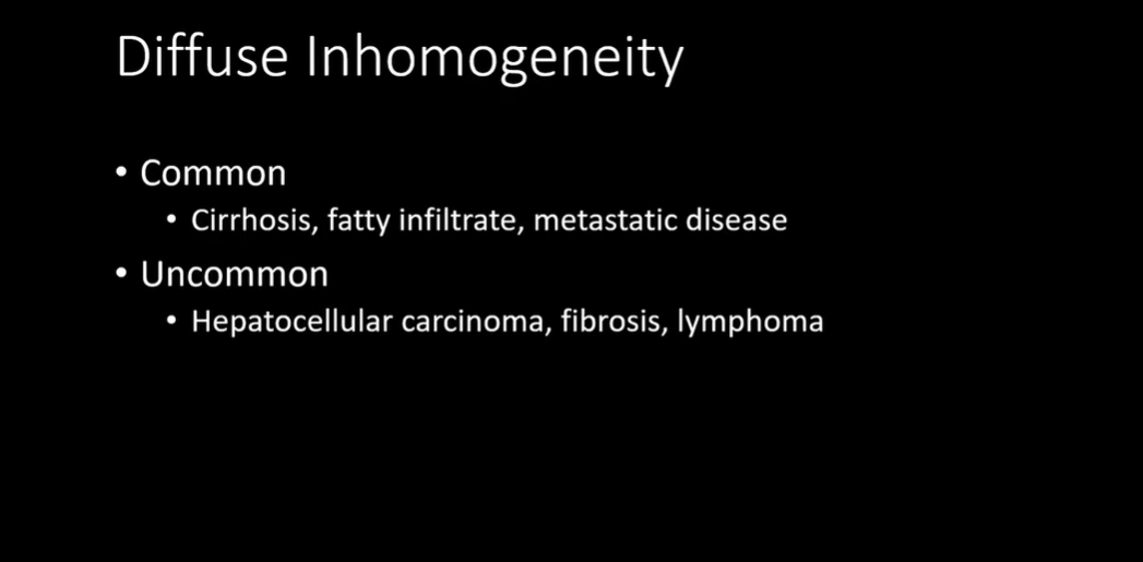
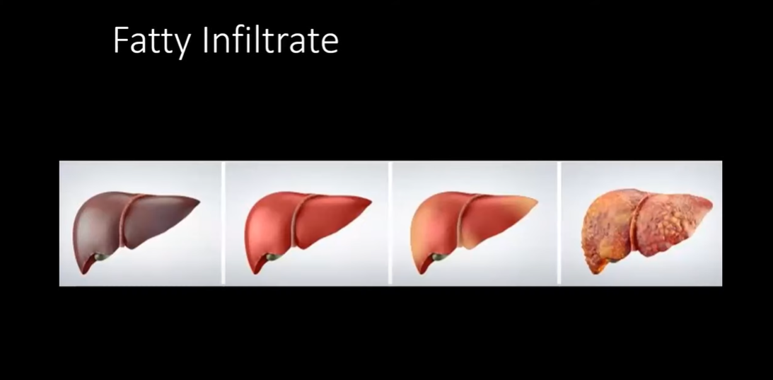
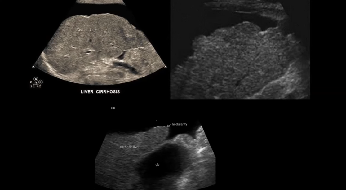
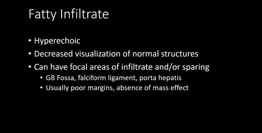
16:24 Fatty infiltration of the liver:
17:18 Another example of fatty infiltration of the liver:
17:52:
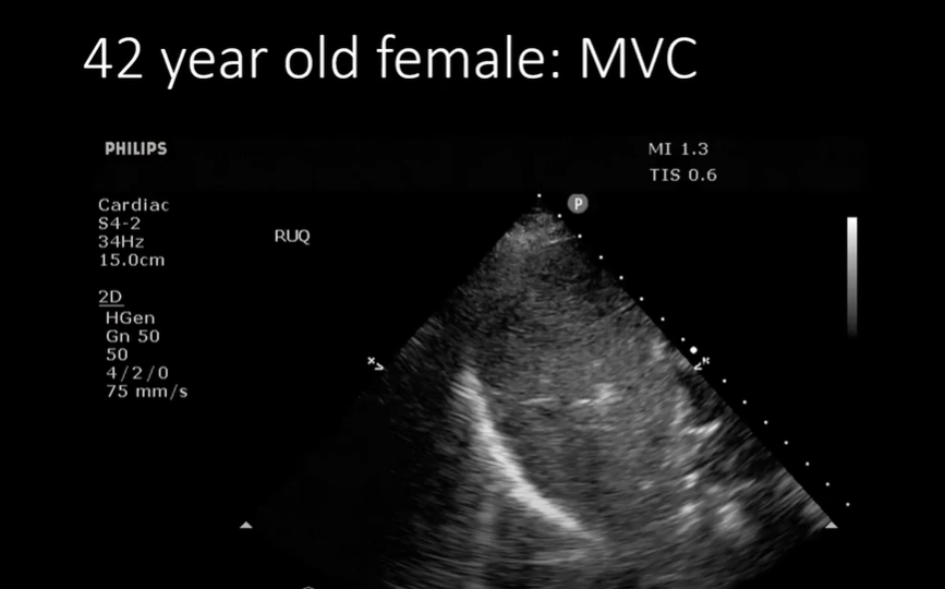
18:15: MVC-So we do the FAST exam.
18:34: And scanning through the liver-what is this round thing-it doesn’t look like what we would see with trauma.
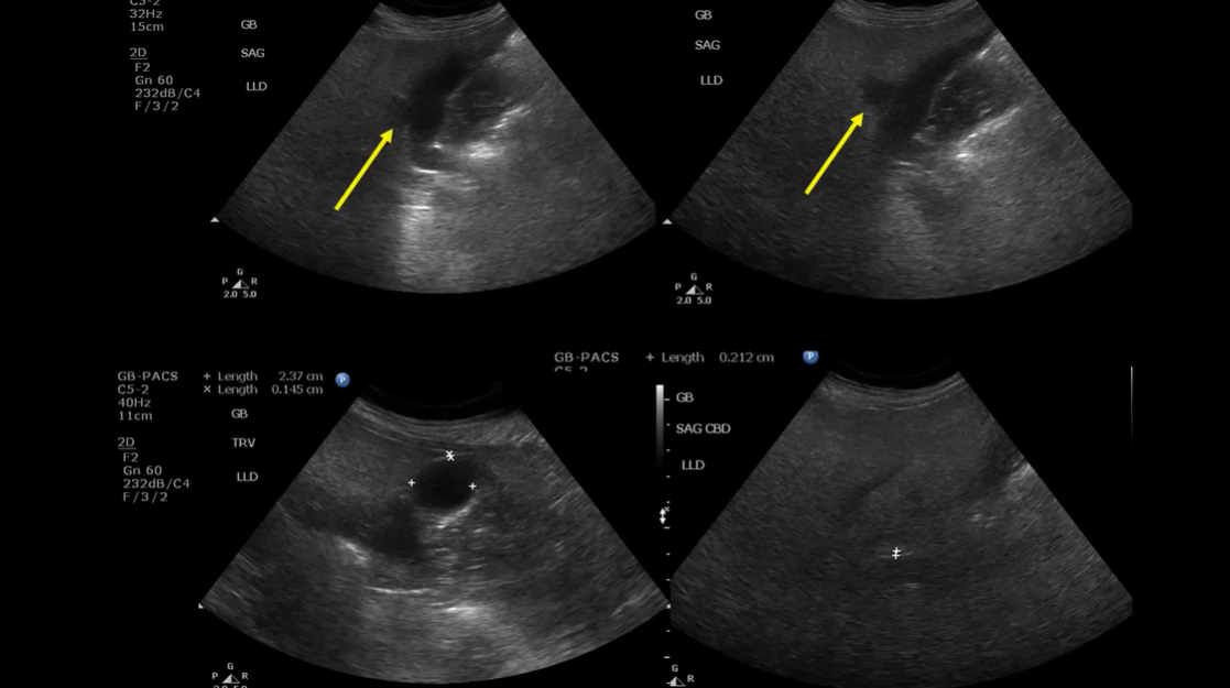
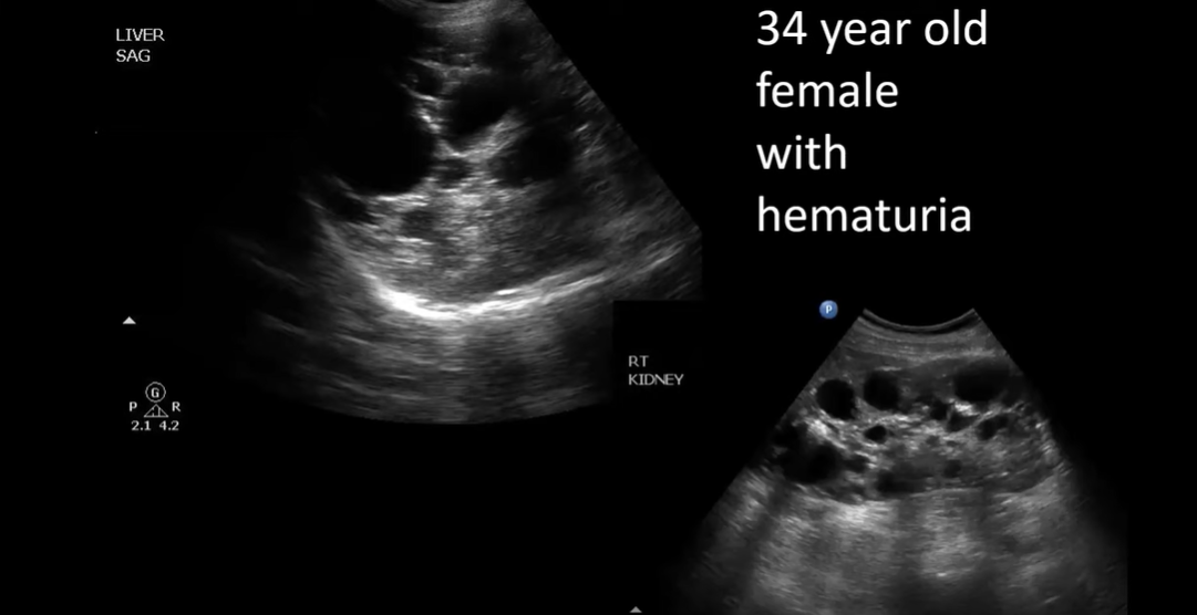
18:53 Another case-some cysts:
18:58 And then wetakea look at the kidneys and we see:
19:29
19:38 Next case:
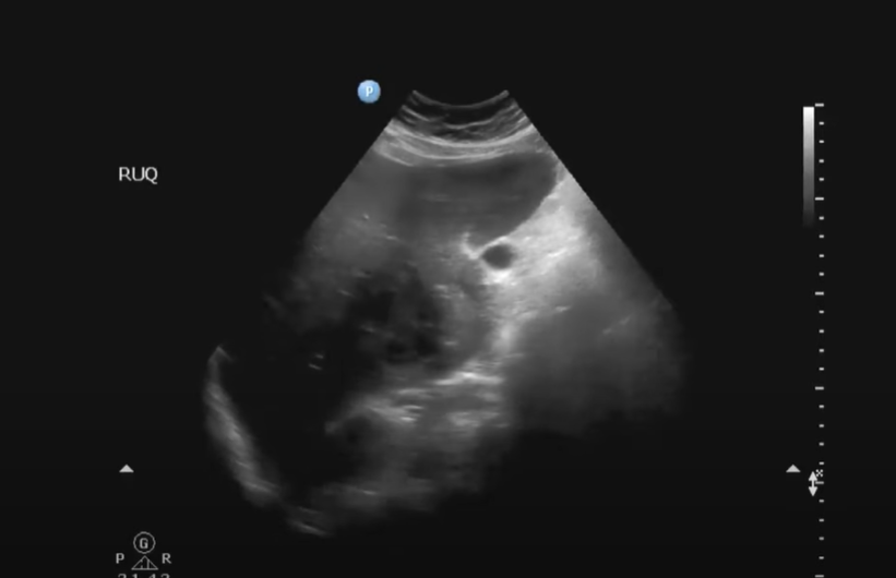
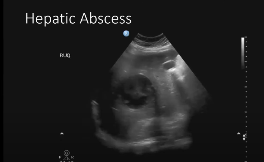
19:49 And so we take a look at his liver.
20:05 He is febrile and there are echos in the mass and so we guess hepatic abscess and it is.
20:12 And if it looks like this you might be thinking, wow this looks like a whole bunch of baby cysts in there. And perhaps this is a hydatid cyst.
21:00
21:12:
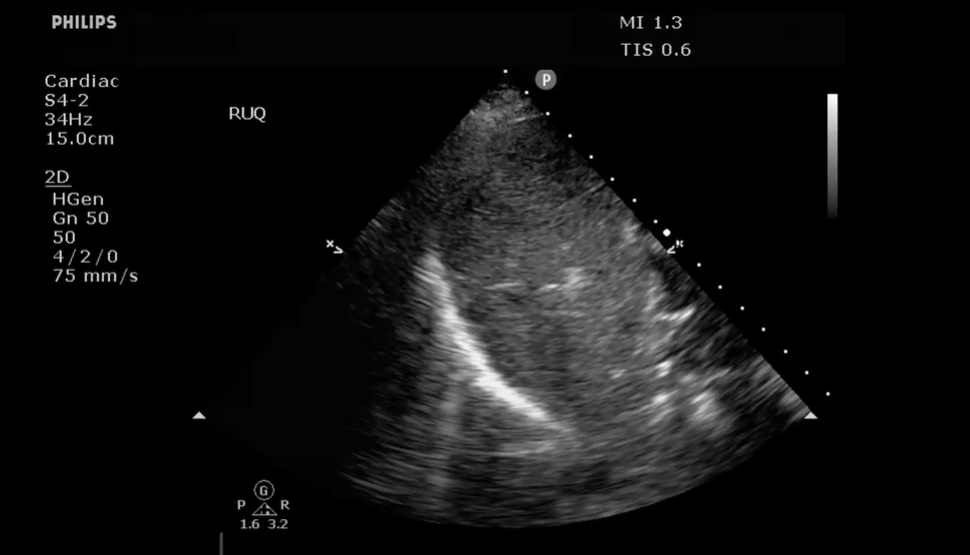
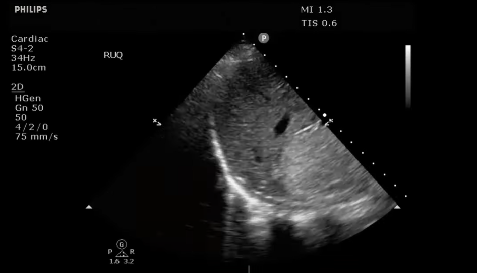
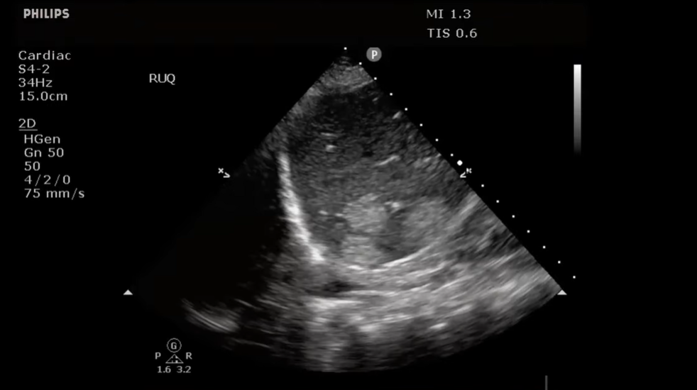
21:46 So now back to our motor vehicle accident patient:
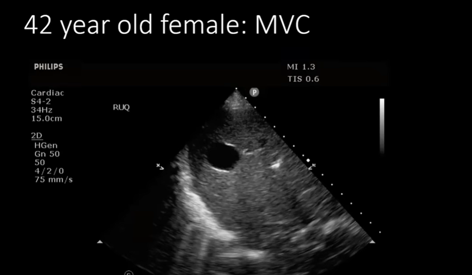
21:55 And you see this:
21:58 And we see these. We first saw the anechoic and now we see hyperechoic lesions in the liver.:
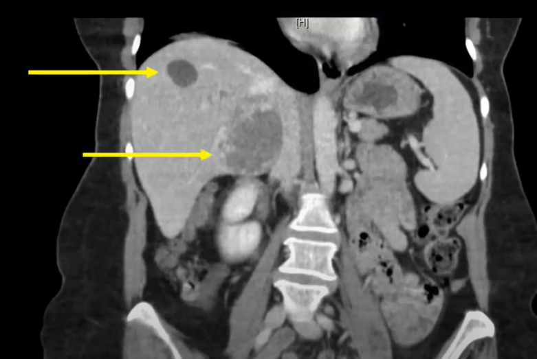
22:06: So on CT we see the anechoic cyst (top arrow) and the hypo-echoic lesion (bottom arrow): The bottom lesion was hyperechoic on ultrasound and hypodense on CT.
22:22:
22:26:
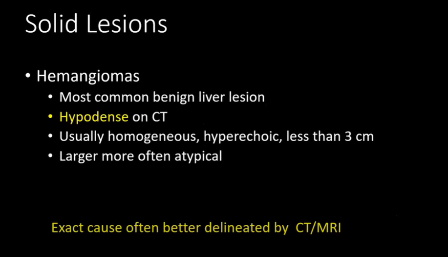
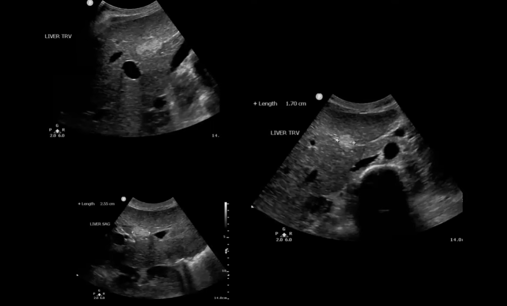
23:01 This is an example of several hemangiomas
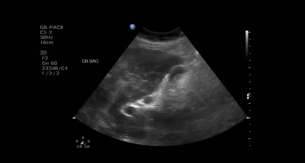
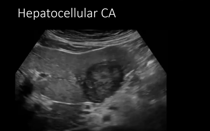
23:12 This is a patient who was placed in our clinical decision-making unit with chest pain, epigastric pain. It wasn’t really clear so I thought I would take a look at the gallblader [which was okay].
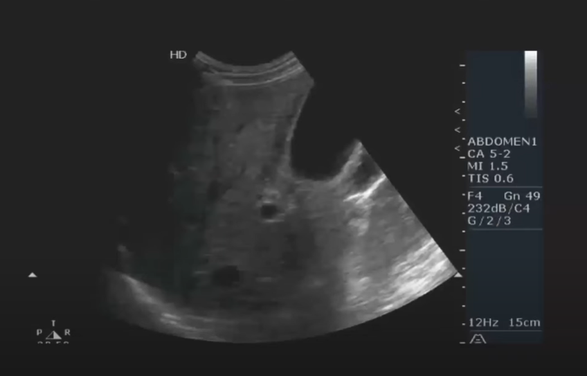
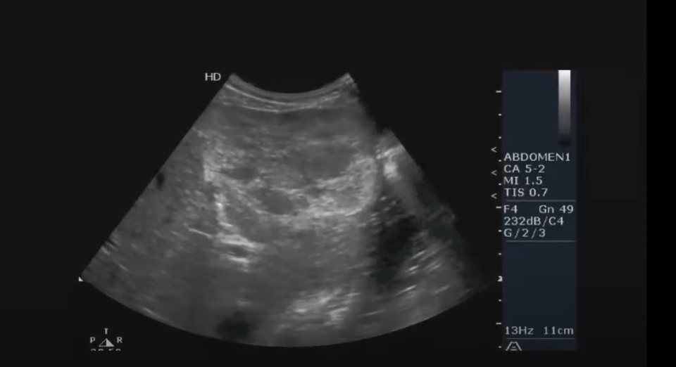
23:25 But sweeping through the liver, you can see anterior [orsuperior to the gallbladder in this orientation] you can see this kind of ratty looking area. Like there is a mass there. It’s not really hyperechoic [but rather] it is kind of mixed.
.
24:07 [So I asked the radiologist what was the next imaging step for this lesion below]:
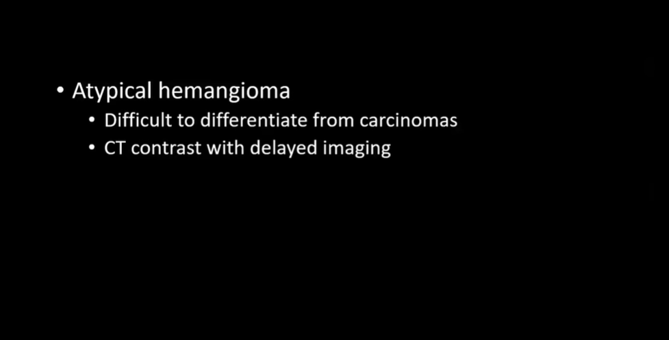
And he said it was either a hemangioma or a hepatocellular carcinoma.
You want to get CT imaging with delayed [contrast] imaging. And if it fills up with contrast, it is a hemangiomma.
It turned out to be a hemangioma.
24:41:
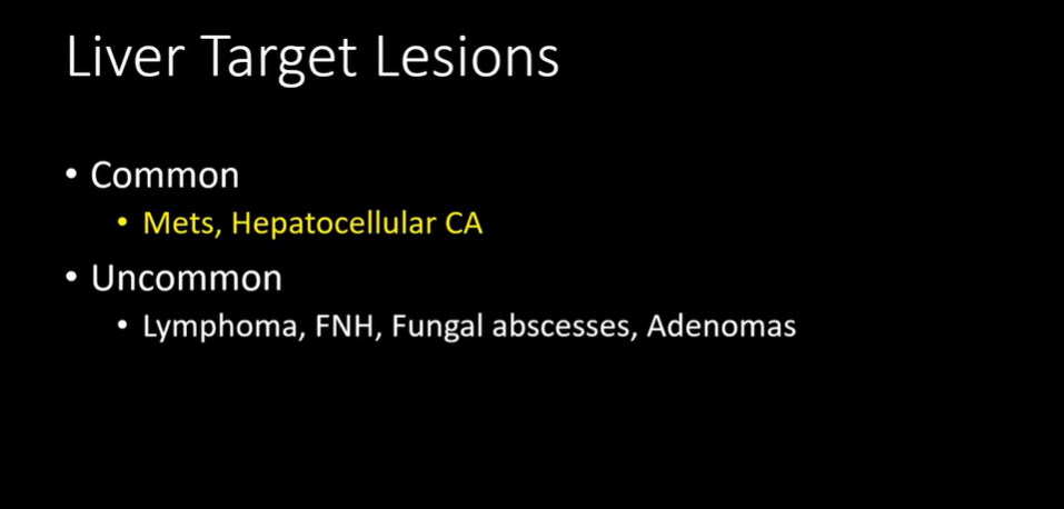
24:5o: These are not very common.
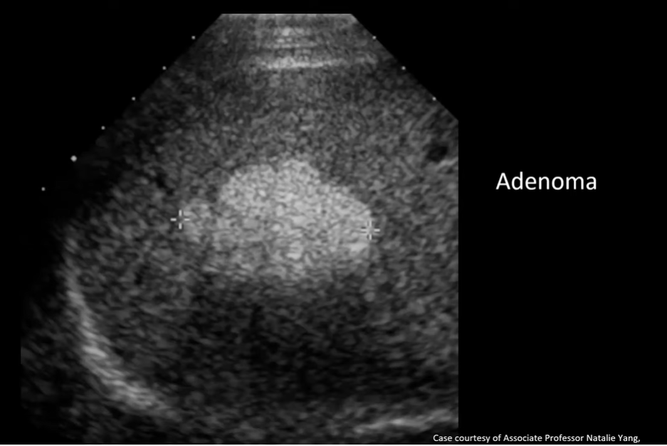
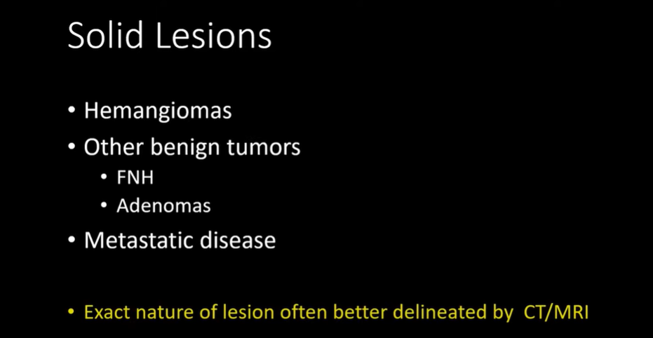
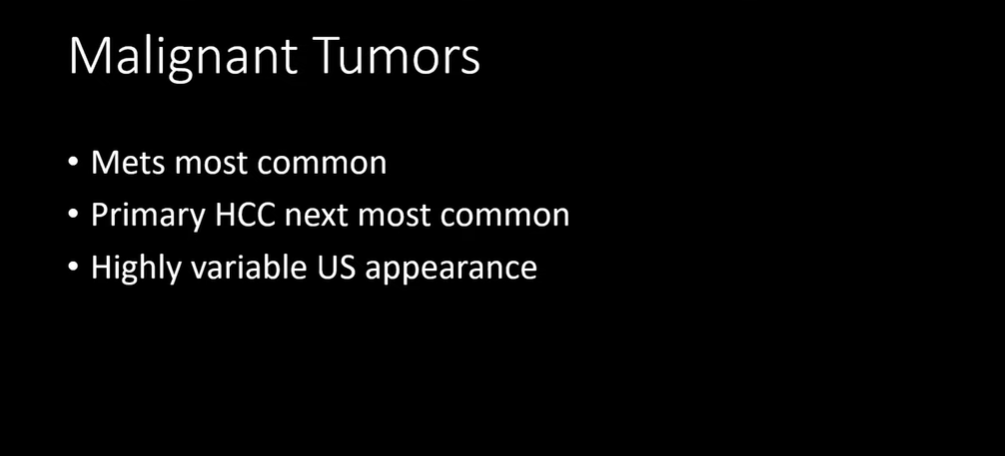
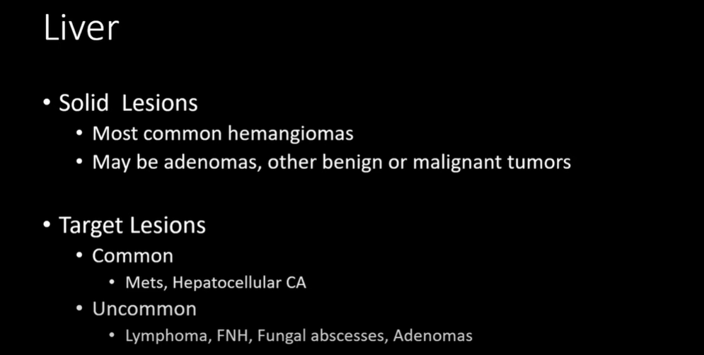
25:33Hemangiomas are far and away the most common solid lesions. They are benign tumors.
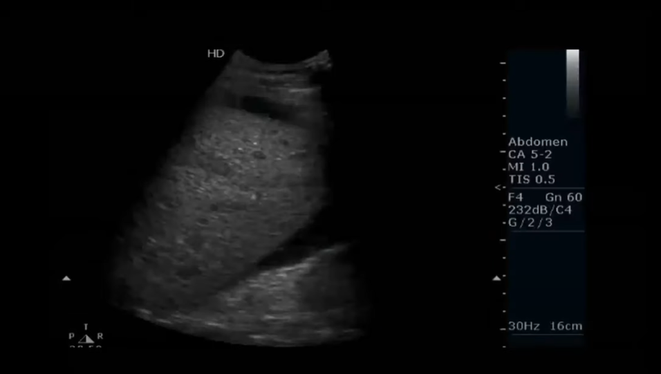
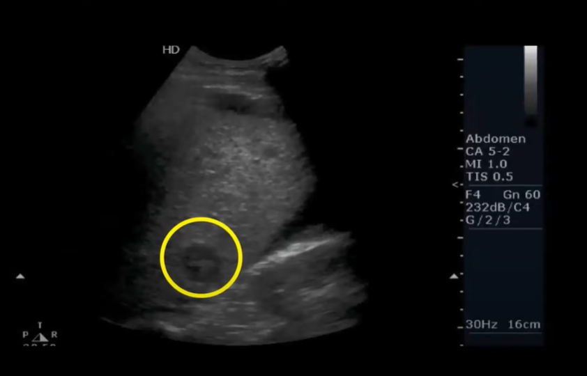
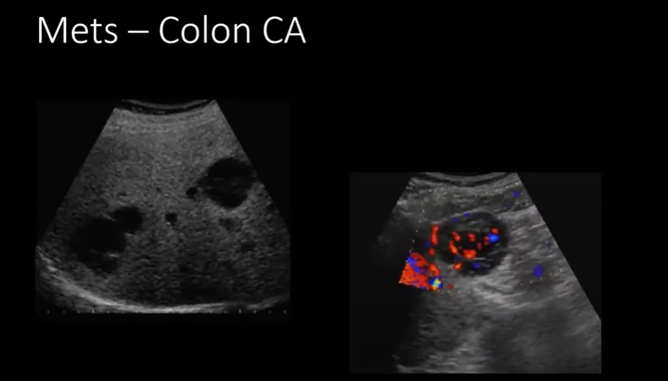
26:18: There is free fluid around this liver and it is not really homogeneous. And it has these lesions. And going through the scan . . .
26:37 . . .you can see this target lesion at the end of the scan.
26:47
27:00
27:48
28:10
28:22
28:29
28:44
29:44
29:45
30-16
30:53
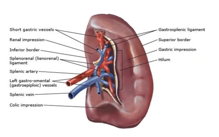
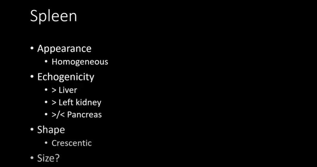
31:00 Anatomy of the spleen.
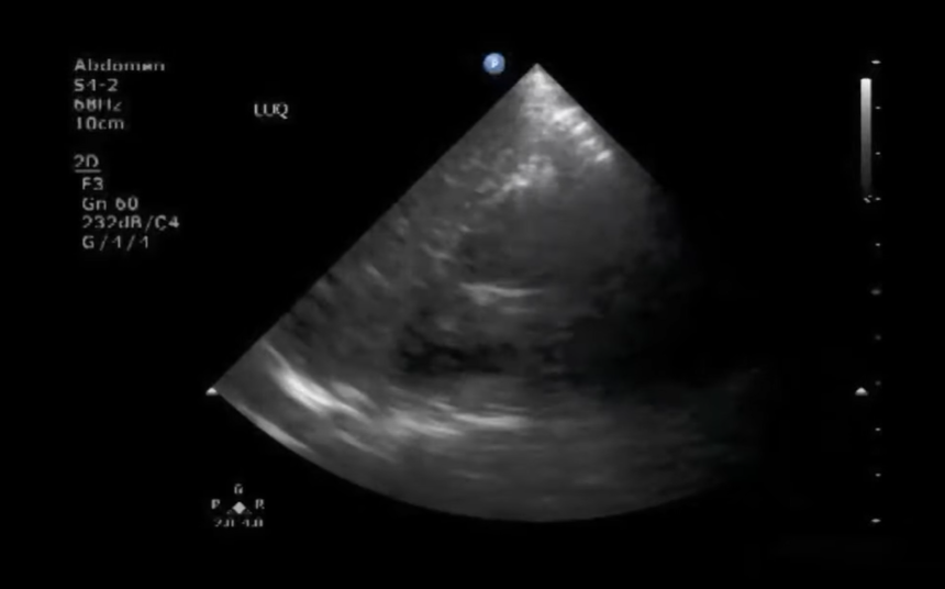
31:48 Scanning through the spleen
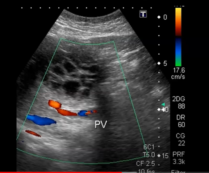
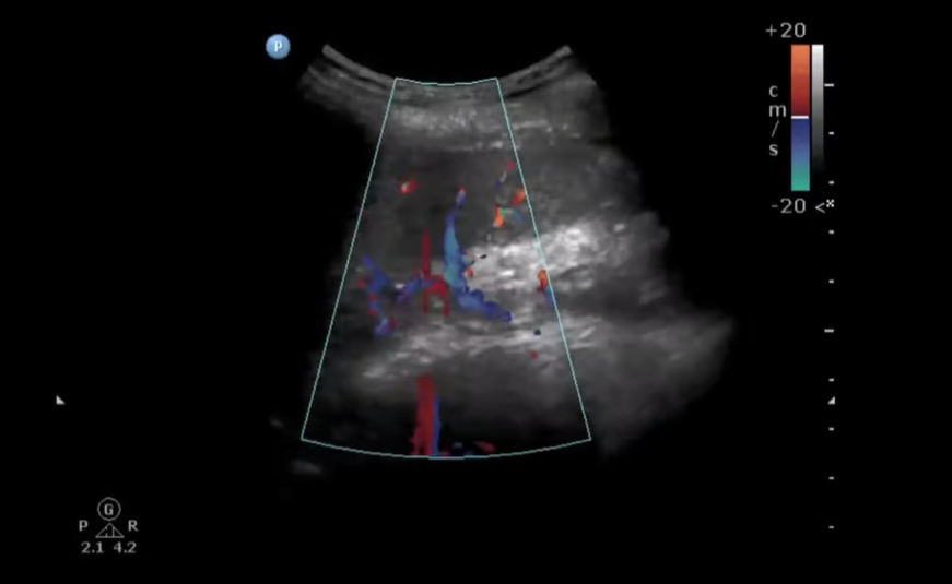
32:01 The splenic hilum with color
32:45
t
66
t
67
t
68
t
69
t
70
t
71