Note to myself: I’ve reviewed this resource before in Links To And Excerpts From The Curbsiders’ “#306 Bronchiectasis and Non-Tuberculous Mycobacterium” With Links To Additional Imaging Resources. Posted on November 29, 2021 by Tom Wade MD
I review the podcast and show notes again because it helps me reinforce the topic
In the above post, I included some imaging resources relevant to the topic
- Imaging protocols for CT chest: A recommendation [PubMed Abstract] [Full-Text HTML] [Full-Text PDF]. Indian J Radiol Imaging. Jul-Sep 2019;29(3):236-246
- Diagnostic Imaging Pathways – Dyspnoea (Chronic). Date Reviewed: 2012
- Diagnostic Imaging Pathways – Bronchiectasis. Date reviewed: May 2018.
- Bronchiectasis (summary)
Last revised by Dr Jeremy Jones◉ on 09 May 2018 from Radiopaedia - High-resolution CT
Last revised by Dr Daniel J Bell◉ on 27 May 2020 from Radiopaedia - HRCT chest (protocol)
Last revised by Travis Fahrenhorst-Jones◉ on 07 Apr 2022 from Radiopaedia - Causes of air trapping on high-resolution CT chest (mnemonic)
Last revised by Dr Owen Kang◉ on 26 Apr 2020 from Radiopaedia - HRCT chest – expiration (protocol)
Last revised by Andrew Murphy◉ on 07 Apr 2022 from Radiopaedia.
In this post, I link to and excerpt from The Curbsiders‘ [Link is to the full episode list] #306 Bronchiectasis and NTM: You can’t get enough sputum! NOVEMBER 22, 2021 By CYRUS ASKIN.
All that follows is from the above resource:
BRONCHIECTASIS INSIGHTS FROM THE WORLD RENOWNED PULMONOLOGIST
DR. PJ MCSHANE, MD
Take a deep breath and inhale high-yield knowledge molecules about the perplexing conditions known as bronchiectasis and NTM (nontuberculous mycobacteria)! Learn the pitfalls and pearls of diagnosis and management from one of the world’s experts, the great Dr. PJ McShane from UT Health – East Texas!
Bronchiectasis Pearls
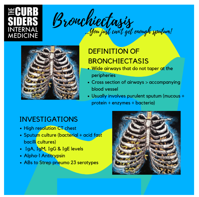
- Bronchiectasis definition: airway widening on CT imaging (larger airway than accompanying vessel); symptoms: usually include purulent sputum, dyspnea/exercise intolerance, cough and potentially malaise and/or hemoptysis.
- Consider eval for bronchiectasis (CT chest) for patients treated for “non-resolving” pneumonia/COPD exacerbations, or, those with obstructive PFTs without a smoking history- especially those encountered in their 5th or 6th decade of life.
- Sputum sampling is incredibly helpful to identify pathogenic organisms that, in the event of an exacerbation, can be targeted for treatment.
- In stable bronchiectasis, airway clearance (+/- hypertonic saline + active cycle breathing maneuvers + some form of vibrating positive pressure device) forms the cornerstone of therapy.
- Bronchiectasis exacerbation definition: 48+ hours of worsening symptoms (3 or more of the hallmark symptoms) plus a clinician’s decision to change/add therapy based upon the clinical presentation.
- Non-tuberculosis mycobacteria (NTM) are environmental pathogens that normally don’t cause disease in immunocompetent patients with normal lung architecture, common agents include Mycobacterium Avium Complex (MAC), M. abscessus and M. kansasii.
- When treatment benefits outweigh risks/adverse events, treatment for MAC in bronchiectasis is a multi-month triple-drug regimen that includes a macrolide (usually azithromycin) + ethambutol + rifamycin.
- In patients that are refractory to the aforementioned treatment, the addition of amikacin liposomal inhalation suspension (“ALIS”) has been demonstrated to increase conversion of positive sputum samples to negative.
Bronchiectasis Show Notes
What is Bronchiectasis? How is it defined?
- Airway + Wide = Bronchi-ectasis!
- Radiographic finding + constellation of symptoms (especially purulent sputum) = the syndrome bronchiectasis, as opposed to the radiographic finding (also called bronchiectasis)
- What is a wide airway? Airway >1x the size of the accompanying vessel in cross-section, on CT scan with a lack of tapering (>1.5x in emphysema) [Shafer 2018]
- Sputum or mucus? Dr. McShane explains that: “Mucus is this wonderfully healthy, slimy, see-through stuff that actually has antimicrobial properties. When mucus gets combined with bacteria, it becomes sputum!”
- How can you describe bronchiectasis to your patient? Dr. McShane tells patients that when mucus becomes sputum, it becomes bogged-down and heavy, and difficult for patients to clear. She further explains that, even though the airways are wide, they don’t work as well and sputum cannot easily be moved out, resulting in worsening infections.
- Strategy for explaining the benefit of airway clearance (see more below): Dr. McShane says that it is similar to sending the police through a broken-down building to ensure squatters don’t take up residence.
So aside from the imaging, what symptoms/history might suggest bronchiectasis?
- Dr. McShane suggests that any patient who requires more than two courses of antibiotics in a year for a “respiratory infection” should get a work up for bronchiectasis, as well as non-smokers with obstructive PFTs.
- She further mentions that dyspnea, especially in association with cough production of sputum, should prompt further work up for bronchiectasis, with CT imaging
Why should we care about bronchiectasis?
- Bronchiectasis can be a major source for morbidity and mortality!
- Prevalence is increasing steadily [O’Donnell 2018]
- US: not a “rare diagnosis,” especially among the elderly (>age 70) where as many as 600/100,000 patients may have bronchiectasis [Akasmit 2017]
- UK: ~ 500/100,000 individuals, all-comers, found via primary care registries [Hill 2017]
- Germany: numbers in between the US and UK [Ringshausen 2015]
- Dr. McShane reminds us that young patients with bronchiectasis, while less common, often have a more complicated course due to their life expectancy at time of diagnosis
Now that bronchiectasis is diagnosed, what to do next?
Thorough history & further evaluation for underlying etiology!
- Ask about systemic syndromes, with concern for connective tissue diseases like rheumatoid arthritis or Sjogren Syndrome [Kelly 2019]
- Asthma symptoms? Consider getting an Immunoglobulin E level [Polverino 2018]
- Family history of lung disease? Emphysematous changes on CT? Consider getting an Alpha-1-antitrypsin level (Cazzola 2020)
- High resolution CTs can identify finer details that may be missed in CTs where the “cuts” are “thicker”, such as tree-in-bud opacities which could be seen in nontuberculous mycobacteria (NTM) infection (Anjos 2020)
- Upper-lobe bronchiectasis can be seen in Cystic Fibrosis, and if this is seen a fertility history can be very helpful (Wielputz 2016)
- Lower-lobe bronchiectasis can be seen in Primary Ciliary Dyskinesia, should prompt questions regarding childhood infections, sinus infections and ear infections as well as fertility history (Knowles 2016)
- Consider checking Immunoglobulins (IgG + subtypes, IgA and IgM), and antibodies to the Strep Pneumonia 23 serotypes (confirming successful immunization) – especially if the patient is getting recurrent infections – can be helpful, and potentially impact therapy (McShane 2021)
- Check sputum early and often: The more the better, per Dr. McShane! Either ask the patient to submit sputum samples or arrange for sputum induction if they are unable to produce sputum spontaneously (or wait for the next exacerbation to get sputum…). Sputum cultures will allow for antimicrobial targeting for future exacerbations. Some patients will grow pathogenic gram-negative infections (P. aeruginosa, S. maltophilia), in some cases this may require nebulized antibiotics (Rubin 2008)
- With the exception of gentamicin (per one, relatively small, single center study in the UK, none of the inhaled antibiotics have consistently proven to reduce exacerbation frequency or definitively improve quality of life (QOL) (Murray 2011)
- Experts point out that the reason for this is likely due to the heterogeneity of bronchiectasis, the variability of baseline maintenance therapy in subjects enrolled in the trials, and use quality of life tools that don’t accurately capture QOL specifically in bronchiectasis . Most of the trials reveal important signals that support the use of inhaled antibiotics and most experts acknowledge that there is a role for this therapy is some bronchiectasis patients. (Antoniu 2011)
- Antibiotics that are commonly compounded and used for inhalation are: colistin, amikacin, tobramycin and aztreonam
- Dr. McShane suggests they probably help “to some degree” and should be used in the management of patients with bronchiectasis who have pathogens identified on sputum culture – especially in patients with high disease burden, an referral to pulmonary for initiation and management is definitely the way to go
- Sputum is also helpful for identifying NTM isolates, which results in an entirely different treatment approach, therefore, sputum should be sent for both respiratory cultures and for acid-fast culture (Park 2015)
Airway Clearance: The Therapeutic Cornerstone
- Upon diagnosis of bronchiectasis, Dr. McShane always starts her patients on airway clearance
- Airway clearance: nebulized hypertonic saline (7% or 3% if 7% is not tolerated) + active breathing (active-cycle breathing) + oscillatory/positive pressure device (ex: Aerobika™, Acapella™) (O’Neill 2018, Lewis 2012)
- Dr. McShane does not recommend N-Acetylcystine given its foul-smell and possible irritant effect, an warns us against using DNAse in non-CF bronchiectasis as it can actually cause harm (Tarrant 2017)
- Dr. Askin mentions that in his practice, he often co-administers the hypertonic saline with bronchodilator (albuterol, levalbuterol), but per Dr. McShane adding the bronchodilator is not always necessary
- Why? Minimal data, but some suggestion of improvement in cough (certainly in NTM) and possibly quality of life (Basavaraj 2020, Basavaraj 2017)
- Nebulized hypertonic saline has been shown to objectively improve lung function in CF bronchiectasis (Wark 2018)
- Theoretically, airway clearance may slow progression of bronchiectasis which may be why it can be helpful prior to the point when frank sputum production is noted
- In regards to active-cycle breathing and other airway clearance basics, Dr. McShane refers patients to the Bronchiectasis Toolbox for educational resources
- Bronchiectasis is irreversible, thus, airway clearance must be done indefinitely
Long Term Antibiotics and Bronchiectasis
- While both inhaled and oral antibiotics in bronchiectasis have not been the panacea we hoped for, they are often appropriate in certain cases such as P. aeruginosa as discussed above
- An exception to this is Azithromycin, recommended in many cases of bronchiectasis for its immunomodulatory effect, not for treatment of a particular pathogen (Kanoh 2010)
- However, in bronchiectasis with NTM – particularly MAC which we will discuss shortly, Azithromycin is a critical antimycobacterial agent with effectiveness demonstrated in at least 3 separate studies (Tartaglione 1997, McShane 2015). Therefore, be cautious with use of azithromycin if NTM is present, as it can easily lead to NTM macrolide-resistance, resulting in NTM that becomes incredibly difficult to treat (Doucet-Populaire 2002)
- That is why sputum sampling is so important, and checking for both bacterial and mycobacterial pathogens is of paramount importance
Bronchiectasis Exacerbations
- ATS definition: “a deterioration in local symptoms (cough, increased sputum volume or change of viscosity, increased sputum purulence with or without increasing wheeze, dyspnoea, haemoptysis) and/or systemic upset” (Pasteur 2010)
- The above definition has evolved to be more granular: “A person with bronchiectasis with a deterioration in three or more of the following key symptoms for at least 48 h: cough; sputum volume and/or consistency; sputum purulence; breathlessness and/or exercise tolerance; fatigue and/or malaise; haemoptysis AND a clinician determines that a change in bronchiectasis treatment is required” (Hill 2017)
- Dr. McShane informs us that having sputum “on the books” prior to an exacerbation, allows the clinician to review the data and select an appropriate antibiotic that should be tailored to the patient
- Expert opinion suggests a week of therapy is reasonable to mild cases, however, for P. aeruginosa or more complicated cases, 2-3 weeks may be necessary – in addition to airway clearance which should be increased if possible
- Caveats from Dr. McShane include not using steroids unless otherwise indicated (ex: asthma exacerbation) and being very cautious if hemoptysis is noted – increasing airway clearance could worsen this and thus, in the presence of hemoptysis a rigorously adherent patient may need to back off their airway clearance even during an exacerbation
Non-Tuberculous Mycobacterium (NTM): Pitfalls & Pearls
- Environmental organisms, over 250 species (McShane 2015)
- Most common pathogens include: Mycobacterium Avium Complex (MAC), M. abscessus and M. kansasii
- MAC is most common making up 80% of NTM identified in patients
- Huge range of disease severity based on numerous factors to include host factors, mycobacterial burden and the specific species in question
- Dr. McShane recommended that primary care providers refer patients with NTM to NTM treatment centers to ensure they receive appropriate treatment and to help our epidemiological understanding of the disease*
* Refer to the National Bronchiectasis and NTM Research Registry website which lists referral centers.
MAC specifics (2020 NTM Guidelines & Dr. McShane’s expert opinion)
- MAC in the sputum is not always pathogenic…
- Diagnosis of MAC infection requires (1) radiologic findings (bronchiectasis and tree-in-bud opacities, etc), (2), two positive sputum samples OR one from BAL, (3) symptoms consistent with MAC infection (refractory weight loss, poor appetite, very fatigued)
- Treatment: three drug / synergistic therapy with a macrolide (azithromycin or clarithromycin) + ethambutol + rifamycin (rifabutin or rifampin) until sputum converts to negative, and then for 12 months after sputum is negative, with monthly sputum sampling
- Azithromycin, per the 2020 guidelines, is the preferred macrolide
- These guidelines also suggest three-times-weekly therapy is far better tolerated and equally efficacious for nodular bronchiectasis without significant concurrent emphysematous disease and/or cavitary disease (with CBC and CMP testing once monthly to check for adverse reactions in addition to eye and vision screening)
- One of the biggest updates from the 2020 guidelines is that, for cases where a patient fails to clear their sputum of MAC despite 6 months of appropriate triple-drug therapy, amikacin liposomal inhalation suspension should be added as a first drug with continued monthly sputum collection
- Amikacin liposomal inhalation suspension is the only FDA approved therapy specifically for MAC with data coming out of the CONVERT trial (Griffith 2018)
Resources for more on NTM & Bronchiectasis curated by Dr. Leah Witt:
Non–Cystic Fibrosis Bronchiectasis | American Journal of Respiratory and Critical Care Medicine
Bronchiectasis Review – Nature
Bronchiectasis in a Diverse US Population: Effects of Ethnicity on Etiology and Sputum Culture
Pulmonary Disease Due to Nontuberculous Mycobacteria: Current State and New Insights
What is Bronchiectasis? (ATS Patient-centered resource)
Treating Bronchiectasis (ATS Patient-centered resource)
Airway Clearance Techniques and Devices (Nat. Jewish Hosp. Patient-centered resource)
The Bronchiectasis Toolbox (an Australian-based, multidisciplinary resource for the diagnosis and management of people with bronchiectasis)