
The following are excerpts from Reference (1) in Resources:
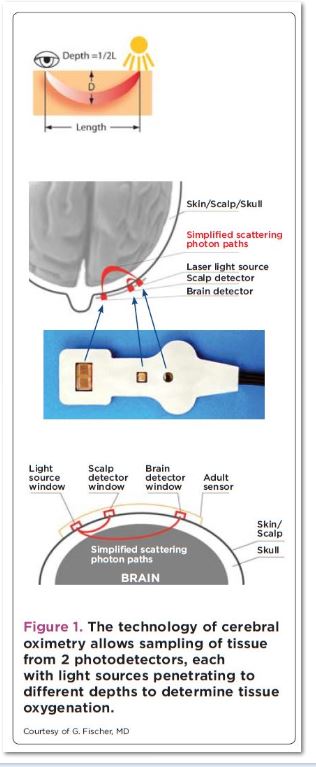
Cerebral oximetry estimates the oxygenation of the regional cortex, an area of the brain that is particularly susceptible to changes in the demand and supply of oxygen, and which has a limited oxygen reserve. Measurement is based on the ability of light to penetrate the skull and to determine hemoglobin oxygenation according to the amount of light absorbed by hemoglobin – – process called near infrared spectroscopy (NIRS). Unlike pulse oximetry (which uses a single sensor), cerebral oximetry with NIRS uses to photodetectors with each light source. The technology allows selective sampling of tissues beyond a specified depth beneath the skin (figure 1). Near field photo detection then can be subtracted from Barfield detection to provide selective measurement of tissue oxygenation adhesive pads applied over the frontal lobes both in the and capture reflected near infrared light passing through the cranial bone to and from the underlying tissue.
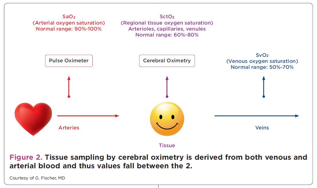
Tissue sampling by cerebral oximetry is mainly from venous (70% to 75%) rather than arterial (25%) blood and is independent of pulsatile flow (figure 2 above). Monitoring is noninvasive and can provide an early warning of decreased oxygen delivery many cardiothoracic and vascular anesthesiologists have adopted the technique to provide continuous intraoperative insight into brain perfusion and oxygenation dynamics.
The FDA has approved four cerebral oximeters: CerOx from Ornim, Equanox from Nonin, Fore-Sight from CASMED, and INVOS by Covidien.
In addition to providing continuous insight into regional oxygenation of the brain, cerebral oximetrymay allow clinicians to use the brain as an index organ that points to the adequacy of tissue perfusion and oxygenation of other vital organs. This concept has received support from multiple clinical outcomes studies (please see references three, seven, and eight, in the article itself).
Conclusion: Evidence suggests that the brain may act as an index organ for how well the vital organs are perfused and oxygenated. Cerebral oximetry has the potential to provide a measurable clinical benefit beyond cardiovascular and thoracic procedures in ever-expanding situations.
The PubMed Abstract Page of reference (2) provides a link to a useful list of other articles on cerebral oximetry.
Resources:
(1) Cerebral Oximetry: Emerging Applications For An Established Technology [Full Text PDF]. Anaesthesiology News, Oct 2012.
(2) Near-Infrared Spectroscopy: The New Must Have Tool in the Intensive Care Unit? [PubMed Abstract] Semin Cardiothorac Vasc Anesth. 2016 May 19. pii: 1089253216644346. [Epub ahead of print]
(3) Cerebral and tissue oximetry [PubMed Abstract] [Full Text HTML] [Full Text PDF]. Best Pract Res Clin Anaesthesiol. 2014 Dec;28(4):429-39. doi: 10.1016/j.bpa.2014.09.002. Epub 2014 Sep 28.


