
This post contains additional resources as well as excerpts from and links to Diagnostic point-of-care ultrasound (POCUS) for gastrointestinal pathology; state of the art from basics to advanced [PubMed Abstract] [Full Text HTML] [Full Text PDF]. World J Emerg Surg. 2018 Oct 15;13:47. doi: 10.1186/s13017-018-0209-y. eCollection 2018.
The article discusses the many abdominal diagnoses in which POCUS may be helpful. Here are some excerpts with additional resources for each sections:
- Background
- Basic Physics
- Examination Techniques
- Free Fluid
- Free Intraperitoneal Air
- Intestinal Obstruction
- Acute Appendicitis
- Epiploic Appendagitis
- Diverticulitis
- Pseudomembranous Colitis
- Intestinal Tuberculosis and Crohn’s Disease
- Colonic Tumors
- Pocus Training
- Conclusions
Background
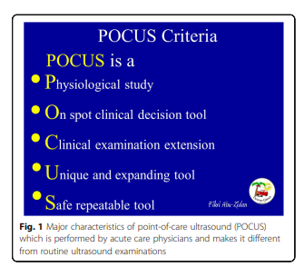
Over the last 30 years, we have realized that POCUS is completely different from the routine radiological studies (Fig. 1). These differences include (1) POCUS is a Physiological study in which the shock status can be evaluated [6, 7], (2) it is an On spot clinical decision tool that helps in critical decision
making in emergency situations within a very short time [8], (3) it is an extension of the Clinical examination and considered as the seventh sense of intra-abdominal inspection [9], (4) it is Unique with expanding indications to study different organs in a systematic approach at the same time [6], and finally, (5) it is Safe and repeatable and
can be used exactly as a stethoscope in the hands of
trained acute care physicians [6].
Basic Physics
Understanding the basic physics of ultrasound and its artefacts is essential to interpret the ultrasound images, to avoid its pitfalls, and to reach an accurate bedside diagnosis. POCUS machines send high-frequency ultrasound waves (2–15 MHz) through their piezoelectric crystals, which are located in the probes, and then receive the reflected waves [10, 11] (Fig. 2).

Examination Techniques
Start here


