In this post I link to the Vimeo Video, The Fastest Way to Diagnose RV Dysfunction by Andre Denault MD and to Doppler Interrogation of the Femoral Vein in the Critically Ill Patient: The Fastest Potential Acoustic Window to Diagnose Right Ventricular Dysfunction? [PubMed Abstract] [Full Text HTML] [Full Text PDF]. Crit Care Explo. 2020 Sep 28;2(10):e0209.
The video is only 24 minutes and closely follows the article . I’d recommend reviewing the video first. In this post I review the video first and then the article.
Here are excerpts from the video:
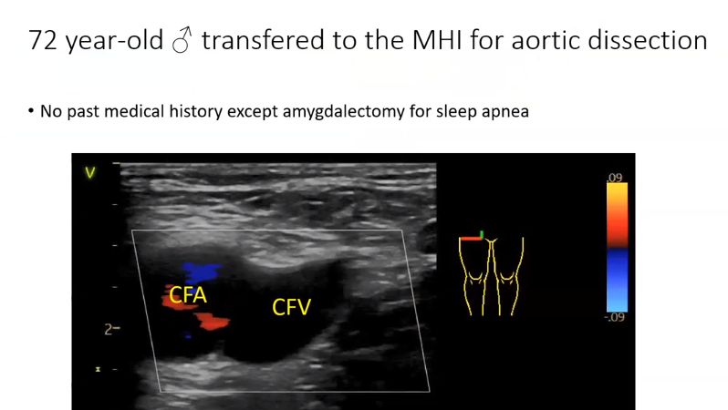
1:17: And in the video below you can see the Common Femoral Vein pulsate.
1:27 And if you use pulse wave doppler, this is what you see. You see a significant reversal of the flow:
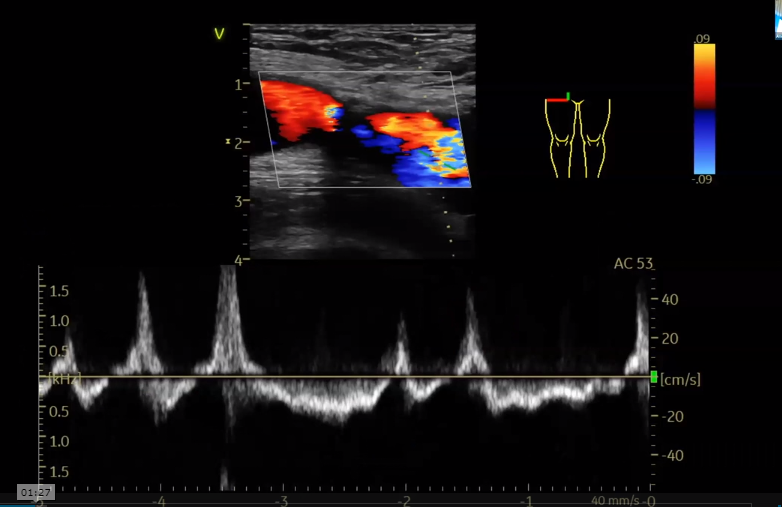
1:35: So the question is how do you feel about that when you this signifcant reversal of flow? Skeptical? Anxious? Well, what is the meaning of a pulsatile venous flow?
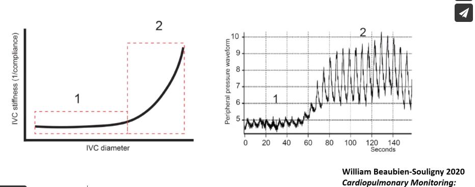
2:04: Basically, when you look at the Inferior Vena Cava stiffness and diameter and you look at the same time at the peripheral pressure wave form*
*Cardiopulmonary Monitoring: Basic Physiology, Tools, and Bedside Management – Was not able to find this citation.
2:18: There is very small little wave form when you have a normal IVC diameter:
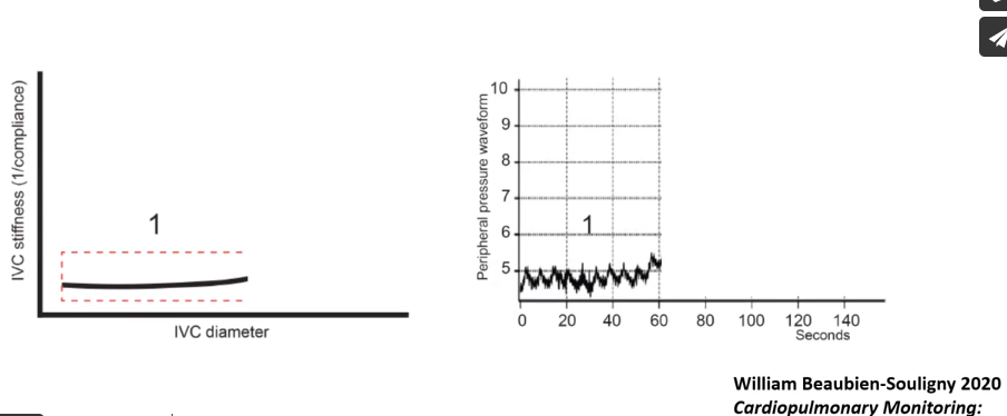
2:25: However, as you increase the diameter and the stiffness, what you will see is the peripheral vein will start to pulsate:
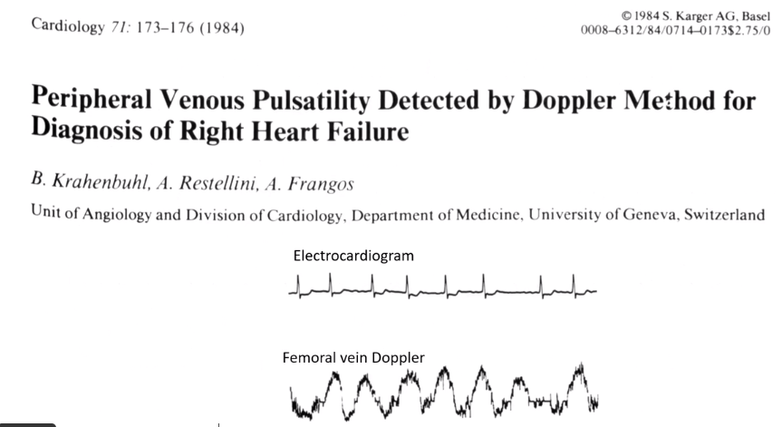
3:40: The normal femoral vein [signal] is continuous with respiratory variation. This is what we call respiratory modulation.
3:50: But if you have cardiac modulation which is a change of the doppler signal with every beat, this is abnormal.
4:00: Cardiac modulation is observed with elevated right atrial pressure, pulmonary embolism, right heart failure, or tricuspid regurgitation.
4:25 below:
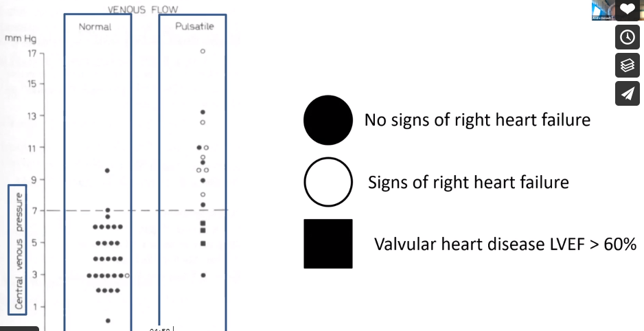
4:30: What you have on this figure [below] is a central venous pressure. So you have those who have a normal venous flow and those who have a pulsatile venous flow.
4:40: And the black dots are those with no clinical signs of heart failure. The white circles are those with signs of right heart failure. And the black squares are those with valvular heart disease with an ejection fraction of more than 60%.
5:00: And the ultrasound at around 7 mm of mercury is the threshold to distinguish those with or without signs of right heart failure.
7:20:
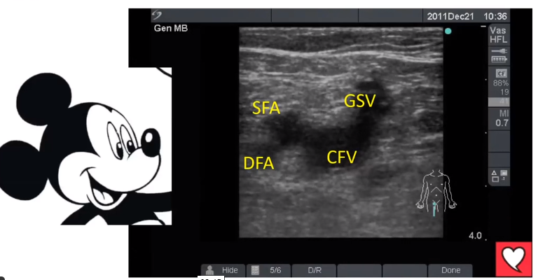
8:15: Compress the vein to make sure you have correctly identified the vein and not the artery because when you have RV failure they will both be very pulsatile:
8:30:

8:36: And this is the anatomy of [the vessels] [And this picture is of the right lower extremity.]
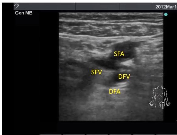
9:00: When you move down a little further, you see this picture [but note that the author is now looking at the left lower extremity]
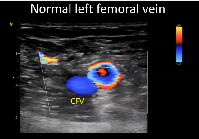
9:05: Once you do your 2D exam, then you do your color doppler.
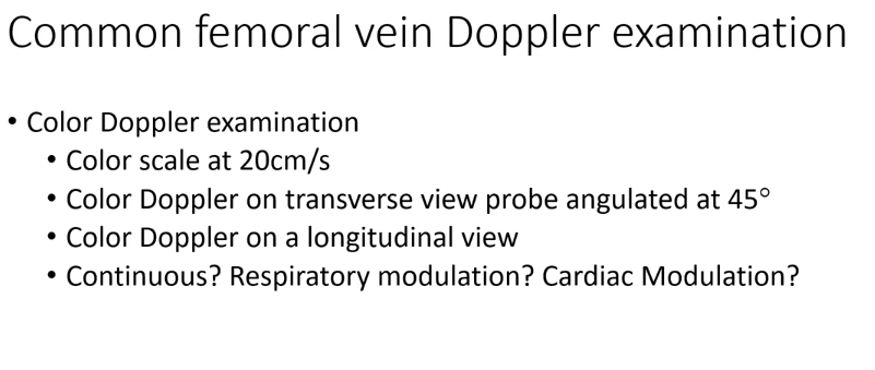
9:45: So you can see that the normal Common Femoral is continuous [always blue] but sometimes a little brighter and this is the respiratory variation.
9:58: This is an abnormal Common Femoral Vein. [The next two photos. The vein is pulsatile – blue, red, blue, red rather than the normal which is continuous blue.]


Note that the CFV above is pulsatile while it should be continuous — just one color, usually blue but it depends on how you have the color scale set.
10:10: Start here.
————————————————————————————————————————-
fig17 is at 11:56 so start here.
And here we see portal vein pulsatility on color flow without any pulse wave doppler. This is abnormal:
fig19
When you look at the arterial and Swann-Ganz signals below, you see:
fig20,
As you pull back, you see there is an overlap with the y-descent:
fig21
And here is the patient’s femoral vein:
fig22
So this next patient, her common femoral vein is continuous. There is a blood clot around the left atrium.And that’s why there is no transmission of the waveform into the femoral vein and so she went right back to surgery:
fig23
Another patient who was unstable with a high lactate. But in this patient the femoral vein is pulsatile (you can see it if you look at the clip, 14:25 – ) and so I was really looking for right ventricular dysfunction. And in the subxiphoid view below you can see that the there is tricuspid regurgitation. This patient improved just with fluid and ino-vasodilators.
fig25 (I skipped fig24)
Start at 15:00
In addition, be sure and review Doppler Interrogation of the Femoral Vein in the Critically Ill Patient: The Fastest Potential Acoustic Window to Diagnose Right Ventricular Dysfunction? [PubMed Abstract] [Full Text HTML] [Full Text PDF]. Crit Care Explo. 2020 Sep 28;2(10):e0209.
Here are excerpts from the article:
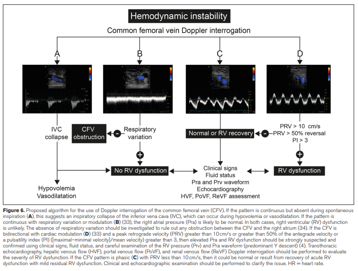
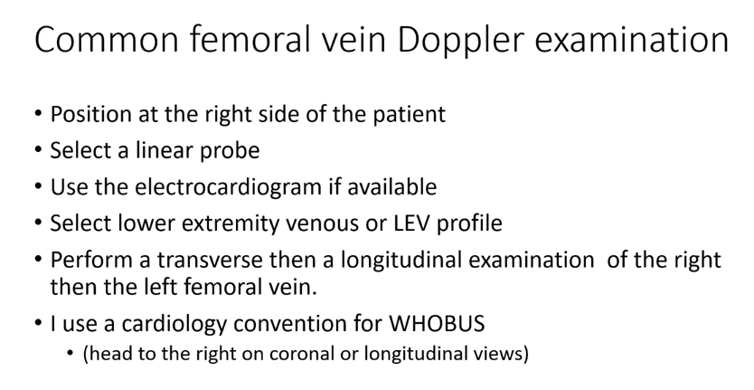
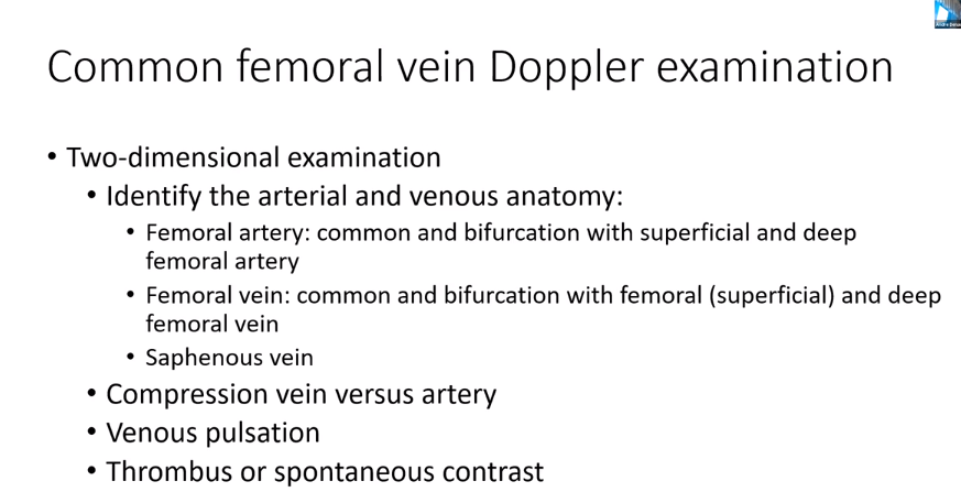
Objectives: To report the use of common femoral vein Doppler interrogation as a simple technique to diagnose right ventricular dysfunction.
Design: Case report.
Setting: Cardiac surgical ICU.
Patients: Postoperative cardiac surgical patients.
Interventions: Common femoral pulsed-wave and color Doppler
examination associated with hepatic, portal, and renal venous
Doppler measurement were obtained in both patients and before and after treatment in patient number 1. In addition, right ventricular pressure waveform examination was obtained in patient number 2.Measurements and Main Results: The technique to obtain common femoral venous Doppler is described. Two cases of patients presenting with right ventricular dysfunction and fluid overload with portal and renal venous congestion in the perioperative period undergoing complex multivalvular cardiac surgery are presented. Hemodynamic waveform monitoring was performed alongside echocardiographic, hepatic, and renal venous flow Doppler assessment, and spectral Doppler profiles of the common femoral veins were examined. Those findings were useful in confirming our diagnosis and guiding our response to treatment. An algorithm was developed and tested on two additional hemodynamically unstable patients.
Conclusions: Doppler examination of the common femoral vein is a simple, fast, and noninvasive technique that could be useful to rule in the presence of right ventricular dysfunction with venous congestion and help guide the management of such patients.
Key Words: bedside ultrasound; common femoral vein; right ventricular dysfunction; venous congestion