
In this post we’ll watch Dr. Turner’s YouTube outstanding video Advanced Pulmonary Ultrasound and then review some of the highlights in notes following the video. And after you have watched the video, be sure and read the 2 articles listed in Resources below. They are the articles on which this video is based.
Advanced Pulmonary Ultrasound
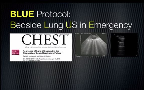
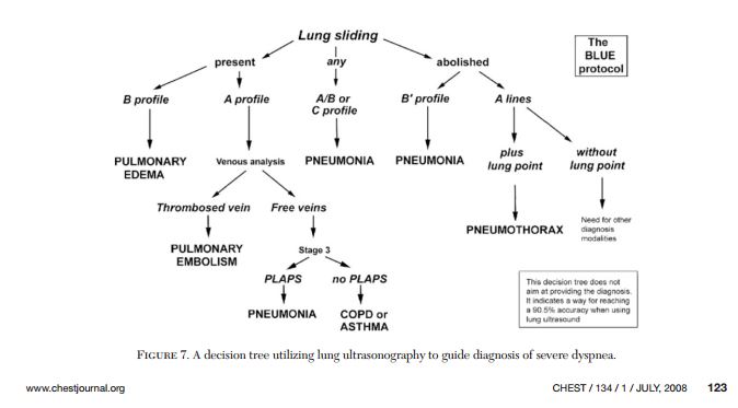
14:30 to 17:50: Below. Dr. Turner discusses the BLUE PROTOCOL (Bedside Ultrasound Scanning In Emergency) (1).
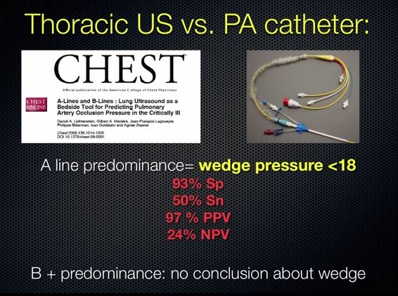
17:50 to 18:39: Below. Dr. Turner discusses the article “A-lines and B-lines: lung ultrasound as a bedside tool for predicting pulmonary artery occlusion pressure in the critically ill.” (2)

18:27: Below. The reason that you can make no conclusion about the wedge pressure when there is B-line predominance is because cardiogenic pulmonary edema (where the wedge is elevated) and non-cardiogenic pulmonary edema (where the wedge is not elevated [as in ARDS]) can look the same.
18:40 to 18:59 : Evidence Based Practice: Literature supports chest ultrasound for the diagnosis of:
- Alveolar-Interstitial Dz (aka edema)
- COPD and RAD
- Pneumothorax
- Pleural Effusion
- Pneumonia
- Hemothorax
- Peripheral Abscess and Tumors
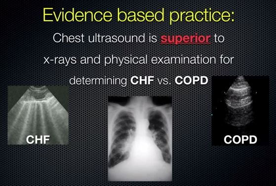
19:00: Evidence Based Practice: Chest ultrasound is superior to x-rays and physical examination for determining CHF vs. COPD.
19:25 Below. The days of the shot gun approach (that is, treating the patient for an acute exacerbation of his CHF and of his COPD) to our patients who have both chronic CHF and COPD who come in short of breath are almost over.
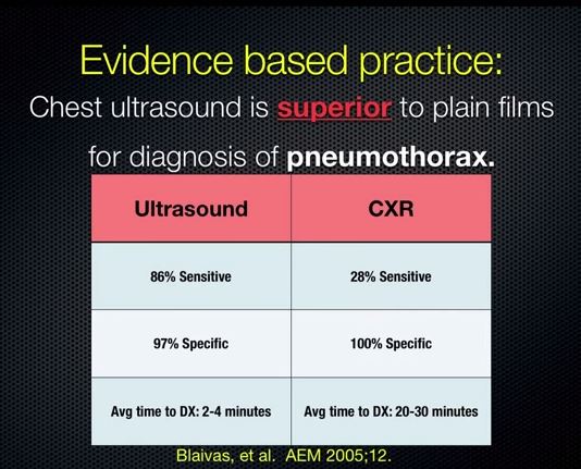
19:48: Below. Evidence Based Practice: Chest ultrasound is superior to plain films for diagnosis of pneumothorax.
20:59: Below.

21:32: Evidence Based Practice: Chest ultrasound improves safety and efficacy of pleural based procedures:
- Chest tube placement
- Thoracentesis
- Biopsy
- Abscess drainage
- Recruitment maneuvers
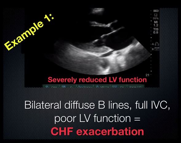
22:27: Below. Shortness of Breath. Example 1.
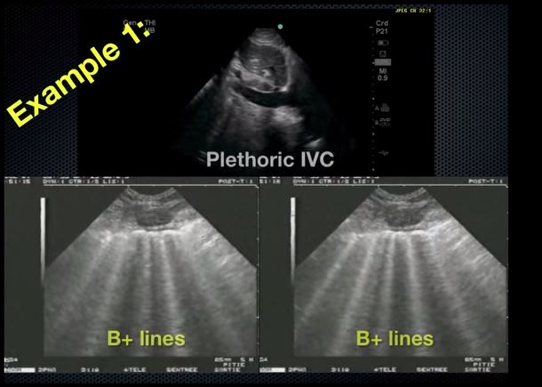
22:42: Below.
23:10: Below. Shortness of Breath. Example 2.
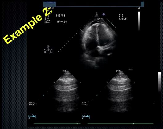
23:26: Below.

23:34 Below.

24:36: Shortness of Breath: Example 3.
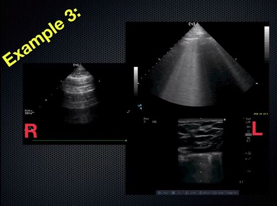
24:20 Below.
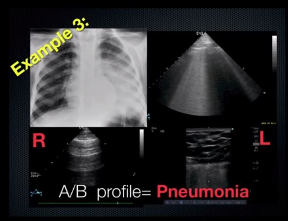
24:58: Below.

Resources:
(1) Relevance of lung ultrasound in the diagnosis of acute respiratory failure: the BLUE protocol.Chest. 2008 Jul;134(1):117-25. doi: 10.1378/chest.07-2800. Epub 2008 Apr 10. [PubMed Abstract] [Full Text HTML] [Full Text PDF]
Correction to [Above] Text: Relevance of Lung Ultrasound in the Diagnosis of Acute Respiratory Failure*: The Blue Protocol FREE TO VIEW
Chest. 2013;144(2):721. doi:10.1378/chest.13-1508. [Correction HTML]: “The article ‘Relevance of Lung Ultrasound in the Diagnosis of Acute Respiratory Failure*: The Blue Protocol’ published in the July 2008 issue of CHEST (2008;134[1]:117-125) contained an error in the text on page 120. The error was in the paragraph that starts with Pulmonary Embolism in the right column under the Results subtitle.” “The correct paragraph reads: Pulmonary Embolism: Pulmonary embolism was observed in 21 patients. Twenty patients had anterior predominant A lines with lung sliding. One had anterior consolidation with absent lung sliding. PLAPS was found in 11 patients. Seventeen patients had venous thrombosis. The article has been corrected.”
(2) A-lines and B-lines: lung ultrasound as a bedside tool for predicting pulmonary artery occlusion pressure in the critically ill. Chest. 2009 Oct;136(4):1014-20. doi: 10.1378/chest.09-0001. [PubMed Abstract] [Full Text HTML] [Full Text PDF]


