Causes of Right Axis Deviation And Of Left Axis Deviation From StatPearls

In this post I link to and excerpt from Electrical Right and Left Axis Deviation, Last Update: January 24, 2021 from StatPearls.

The above article also has an excellent brief review of how to determine the QRS axis.

Here are excerpts:

Electrical Axis Classification

There are five main electrical axis classifications: 

  1. normal axis
  2. left axis deviation (LAD)
  3. right axis deviation (RAD)
  4. extreme axis deviation, and
  5. indeterminate axis

The following axis classifications described are based on adults.

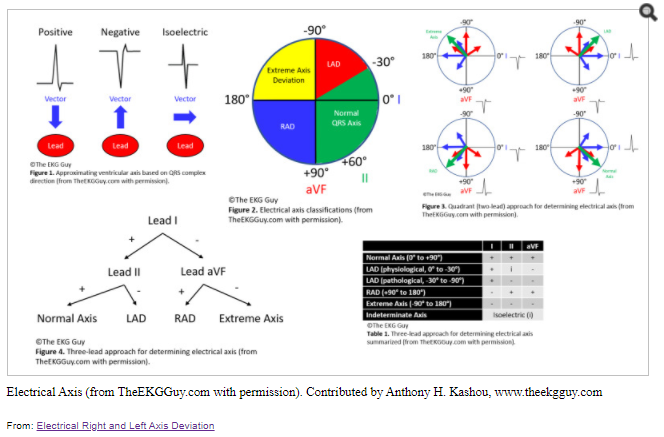
If the QRS axis falls between -30 degrees and -90 degrees, it is considered LAD. In this case, the QRS vector is directed upward and to the left.

If the QRS axis falls between +90 degrees and 180 degrees, or beyond +100 degrees if the adult range is used, then RAD is present. The QRS vector would be directed downward and to the right.

If the QRS axis happens to fall between -90 degrees and 180 degrees, this would be referred to as extreme axis deviation, whereby the ventricular vector is directed upward and to the right.

Lastly, if the QRS complex is isoelectric or equiphasic in all leads with no dominant QRS deflection, it is considered an indeterminate axis. The electrical axis classifications are summarized in Figure 2.

Clinical Significance

Causes of LAD include: 

  • Normal variation (physiologic, often age-related change)
  • Left ventricular hypertrophy
  • Conduction defects: left bundle branch block, left anterior fascicular block
  • Inferior wall myocardial infarction
  • Preexcitation syndromes (e.g., Wolff-Parkinson-White syndrome)
  • Ventricular ectopic rhythms (e.g., ventricular tachycardia)
  • Congenital heart disease (e.g., primum atrial septal defect, endocardial cushion defect)
  • Hyperkalemia
  • Emphysema
  • Mechanical shift, such as with expiration or raised diaphragm (e.g., pregnancy, ascites, abdominal tumor, organomegaly)
  • Pacemaker-generated rhythm or paced rhythm

Causes of RAD include:

  • Normal variation (e.g., children, young adults)
  • Limb-lead reversal (left- and right-arm electrodes)
  • Right ventricular overload syndromes (acute or chronic)
  • Right ventricular hypertrophy
  • Conduction defects: left posterior fascicular block, right bundle branch block
  • Lateral wall myocardial infarction
  • Preexcitation syndromes (e.g., Wolff-Parkinson-White syndrome)
  • Ventricular ectopic rhythms (e.g., ventricular tachycardia)
  • Congenital heart disease (e.g., secundum atrial septal defect)
  • Dextrocardia
  • Left pneumothorax
  • Mechanical shift, such as with inspiration or emphysema
  • Conditions that cause right ventricular strain (e.g., pulmonary embolism, pulmonary stenosis, pulmonary hypertension, chronic lung disease, and resultant cor pulmonale)
This entry was posted in Electrocardiography. Bookmark the permalink.