
As a primary care clinician, I probably don’t need to know even the basics of this subject. And this post features only the basics. This post is really just because I find the subject of medical imaging so interesting.
However, I came across the following article, The Central Vein Sign: Is 1.5 T MRI still relevant for diagnosis of multiple sclerosis? [Full Text HTML] [Full Text PDF]. Practical Neurology, February, 2020. This article got me started on this review post.
Here are excerpts:
MRI and Multiple Sclerosis Diagnosis
The most important paraclinical tool for the diagnosis of multiple sclerosis (MS) is MRI, which is included in all iterations of the McDonald criteria, although the magnetic field strength is not specified. Conventional 1.5T MRI is most frequently used in the diagnostic process and has high sensitivity but lower specificity for the detection of white matter (WM) abnormalities.
Despite technologic advances and updated McDonald criteria improving the accuracy of MS diagnosis, there are complex or atypical cases for which the diagnostic value of 1.5T MRI is limited, especially for nonspecific lesions, very few small lesions, or lesions restricted to the supratentorial area, particularly in the context of comorbid conditions. Differential diagnosis between MS and MS mimics remains challenging, and misdiagnosis is highly prevalent in MS (~20%), leading to treatment delay or unnecessary initiation of DMTs.1,2 Clinicians commonly encounter a clinicoradiologic paradox.
There remains an unmet need for sensitive biomarkers to facilitate accurate diagnosis and guide treatment decisions. Development of high-field (≥3T) and ultrahigh-field MRI (≥7T) can potentially overcome limitations of the 1.5T MRI and further strengthen the role of MRI in the diagnosis and management of MS.
Pros and Cons of High-Field MRI
High- and ultrahigh-field MRI have been widely used in research and are becoming more available in the clinical setting. Although 3T MRI is approved for clinical use, 1.5T MRI remains most available in clinical practice. In 2017, the Food and Drug Administration (FDA) cleared the first 7T-MRI device for limited use in clinical practice, restricted mainly to head and extremity examination.
Conventional 1.5T MRI is limited because of a low signal-to noise ratio (SNR) and modest spatial resolution. High-field MRI has the advantages of a higher SNR and a higher contrast-to-noise ratio (CNR) to provide enhanced spatial resolution up to 100 µm. This increase in spatial resolution improves visualization of small anatomic structures, separation of gray and white matter, and accurate detection of subtle abnormalities, including small lesions.3-7
In ultrahigh-field MRI, susceptibility effects, subtle field perturbations generated by local variations in magnetic properties, are boosted, especially in proximity to tissues with high iron content8 and densely distributed myelinating axons,9 allowing examination of microscopic veins, iron in the brain, microbleeds, and densely myelinated structures (eg, optic radiations).
Central Vein Sign – a Case for >1.5T Field Strength
Early histopathologic studies reported that most demyelinating lesions are centered on small parenchymal veins and this is confirmed by high-field MRI (3T and 7T) using T2-weighted sequences.
The CVS has been observed in all clinical phenotypes of MS, including relapsing and progressive forms of the condition. The CVS has been proposed as an imaging biomarker of great diagnostic value for distinguishing between MS and MS mimics.
The current understanding is that the presence of a CVS can accurately differentiate MS from similar nonMS pathology provided a minimal cut-off between 40% to 50% of lesions with the CVS is reached.10
Diagnosis of Multiple Sclerosis
Given the higher sensitivity of 7T MRI in detecting cortical lesions and the CVS, 7T MRI can play a key role in improving MS diagnostic accuracy. With 7T MRI, cortical lesions, undetectable on 1.5T or 3T MRI, can be seen; this can increase diagnostic yield, now that cortical lesions are included in the MS diagnostic criteria.
Conclusion
What is the future of high- and ultrahigh-field MRI? Is it just a matter of time to find widespread application and use in everyday clinical practice?
Larger clinical studies using 7T MRI for diagnosis and treatment decisions are needed before introducing ultrahigh-field MRI into everyday clinical practice.
Although high-field MRI will be more widely used in research, and there are unparalleled possibilities for the future, 1.5T MRI is still the main diagnostic tool for the MS diagnosis and treatment decisions but its limitations raise the question whether MS patients should be evaluated on high- and ultrahigh-field MRI scanners.
Next, I reviewed this post from DMS Health: How Strong Does Your MRI Magnet Really Need to Be? MAY 16, 2019.
And here are excerpts from the above.
How Strong Does Your MRI Magnet Really Need to Be?
Choosing the right MRI imaging equipment for your hospital or clinic can be a complex decision.
As advances in technology have continued in the MRI field, you have more choices available for machines of higher field strength. Typically, these machines can cost at least twice as much as the commonly used 1.5T machines, so clinics find themselves weighing up the cost-benefit.
One of the big questions people ask is, how strong does that MRI magnet really need to be? Does bigger mean better? Let’s take a closer look at MRI and the relative merits of field strength.
A primer on MRI field strength
“Field strength” refers to the magnetic field strength of the magnet used in the MRI machine. This correlates with signal-to-noise ratio — the stronger the field the stronger the signal. Magnetic field strength is measured in teslas (T) and higher field strength can also equate to faster throughput.
The measurement in tesla is proportional. Therefore, an MRI machine at 3.0T is twice as strong as a machine at 1.5T.
Commercially available MRI scanners for routine clinical use exist from 0.2T to 3.0T, while research facilities currently perform human imaging in fields up to 11.7T. The vast majority of machines in use in a clinical setting are 1.5T (including our mobile fleet at DMS Health).
Channels and coils
One of the most important elements of an MRI to consider is the coils. The coils correlate with how many channels your MRI machine is able to offer. This is through the number of coil elements, so four elements means four channels. A channel refers to the receiver pathway of the MRI system.
More channels mean better image quality and improved speed of acquisition. Many MRI machines in use have 4, 8, 16, even 32 or more channels. On-coil digitization is a technology that is allowing more channels off a lower number of coils, too. This technology can help with the overall price as more elements mean a higher price.
If you’re looking at field strength of an MRI magnet, it’s important to keep in mind that channels play a big role. For example, a unit with a comparatively weaker field strength may have more channels available, which may be more beneficial than one with a higher strength field and less channels.
1.5T vs. 3.0T MRI machines
In comparing 1.5T against 3.0T MRI machines, an understanding of signal-to-noise ratio is important. We explained earlier that the stronger field strength produces a stronger signal — a clearer image may be produced because the stronger signal overcomes background noise. This noise shows up as graininess in images, which will be reduced in 3T as compared to 1.5T machines. It’s like turning up the volume on your music to drown out background noise.
Resolution
Higher field strength also correlates with higher temporal resolution and higher spatial resolution. Higher spatial resolution is helpful in gaining clearer images of small, complex structures, such as detecting nerve root, spinal cord or neuroforaminal pathologies when imaging the spine.
Higher temporal resolution makes for a more efficient MRI scanner. Comparing 3.0T and 1.5T machines, it means that the 3.0T scanner may be able to throughput more patients in the same amount of time with similar image quality to the 1.5T scanner. Of course, in facilities with relatively low-volume scanning needs, throughput may be a negligible factor.
Conclusion
How strong does your MRI magnet really need to be? The answer lies with your individual facility in terms of your needs for highly detailed images and the volumes of patients you need to have scanned. A 1.5T field strength is the most commonly used and is considered appropriate for most clinical needs.
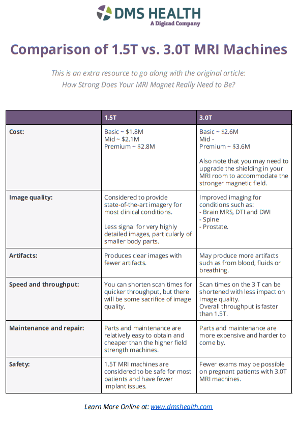
Here is theSummary Sheet, Comparison of 1.5T vs. 3.0T MRI Machines, from DMS Health. – “This is an extra resource to go along with the original article: How Strong Does Your MRI Magnet Really Need to Be?”
And finally I reviewed RF coils: A practical guide for nonphysicists [PubMed Abstract] [Full Text HTML] [Full Text PDF]. J Magn Reson Imaging. 2018 Sep; 48(3): 590–604.
Here are excerpts:
Radiofrequency (RF) coils are an essential MRI hardware component. They directly impact the spatial and temporal resolution, sensitivity, and uniformity in MRI. Advances in RF hardware have resulted in a variety of designs optimized for
specific clinical applications. RF coils are the “antennas” of the MRI system and have two functions: first, to excite the
magnetization by broadcasting the RF power (Tx-Coil) and second to receive the signal from the excited spins (Rx-Coil).
Transmit RF Coils emit magnetic field pulses (B1
1 ) to rotate the net magnetization away from its alignment with the main magnetic field (B0), resulting in a transverse precessing magnetization. Due to the precession around the static main magnetic field, the magnetic flux in the receive RF Coil (B1 ) changes, which generates a current I. This signal is
“picked-up” by an antenna and preamplified, usually mixed down to a lower frequency, digitized, and processed by a
computer to finally reconstruct an image or a spectrum. Transmit and receive functionality can be combined in one RF
Coil (Tx/Rx Coils). This review looks at the fundamental principles of an MRI RF coil from the perspective of clinicians
and MR technicians and summarizes the current advances and developments in technology


