Today, I reviewed, link to and embed Dr. M Usman Javed‘s YouTube video, Echocardiography Standard Protocol | Step by Step | Complete Trans-thoracic Normal Echocardiogram.
Be sure to visit Dr. Usman’s’s website: www.drmusmanjaved.com
All that follows is from today’s outstanding resource and review.
76,974 views Aug 24, 2022 Cardiology
In this video I am going to illustrate the protocol for performing complete and comprehensive transthoracic echocardiography, which meets the current standards of American Society of Echocardiography. I will not be going into a lot of pathological issues here so the protocol presented here is meant as a guideline and does not cover every aspect, such as off axis views. I am also going to talk basic probe position and major structures visible side by side with the protocol steps. So when you follow the protocol you don’t skip anything, you can get additional images according to the pathology found. I personally found that once I could execute the standard protocol flawlessly, I was able to add and refine additional echo scanning skills while deepening my understanding of the purpose of each echo image. So it’s immensely useful to learn basic steps of protocol. We will start with parasternal long axis window followed by short axis, apical and in the end subcostal and suprasternal windows.
0:00
0:25
0:30
0:42
0:52
0:54
1:05
1:13
1:18
1:20
1:25
1:32
1:32
1:36 – 1:54
1:54
1:58
2:03
2:08
2:17
2:21
2:25
2:36
2:40
2:45
2:48
2:55
3:07
3:13
3:22
3:29
2:36 Now take the same three measurements in systole.
2:40
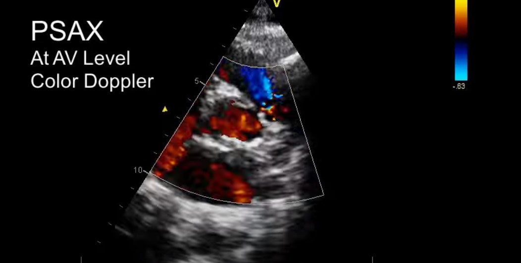
2:42 Zoom aortic valve and . . .
2:47 Then measure . . .
2:55
3:19
3:23
3:27
3:30
3:36 In the next step focus on the tricuspid valve.
3:39
3:45
3:48
3:53
4:00 – 4:26 The speaker whatwecan see of the scallops of the mitral valve
4:28
4:34 Now point the beam more apically
Here is a clearer image of the midcavity from Focused Cardiac Ultrasound for the Nephrologist: The Parasternal Short Axis View. July 22, 2019: [fig47a]
4:43
4:55 Use pointer towards the left shoulder. See YouTube video Echocardiogram How To: Learning The Apical 4 Chamber View from GE HealthCare.
The author in this video uses pointer to the appendix (LV is to the left of the screen) rather than more common pointer towards left shoulder or towards the bed (in which case the LV is on the right of the screen.
From 5:22 – 7:17 The author discusses the views to be taken. Since he is has the LV to the left side of the screen, I have added an additional resource below. But this secion still needs to be carefully reviewed.
Review, also, this link on the apical views: Focused Cardiac Ultrasound for the Nephrologist: The apical window. September 20, 2019.
And finally, review How to obtain: APICAL VIEWS! (Echocardiography) from the echo lady
7:25
7:31
7:49
7:58 Looking for atrial septal defect