Today, I review, link to, and excerpt from Chinese Clinical Oncology‘s “Update on management of pancreatic cancer: a literature review”. [PubMed Abstract] [Full-Text HTML] [Full-Text PDF]. Chin Clin Oncol. 2024 Jun;13(3):41. doi: 10.21037/cco-23-94. Epub 2024 May 17.
There are 126 similar articles in PubMed.
All that follows is from the above resource.
Introduction
Approximately 85–95% of pancreatic cancers are pancreatic ductal adenocarcinomas originating from the exocrine part of the pancreas, and the term “pancreatic cancer” typically denotes this disease entity (1). It is an aggressive malignancy with increasing in incidence and with a high mortality. Globally, pancreatic cancer is currently the 7th highest cause of cancer-related death, and with the current trend, pancreatic cancer is projected to be the 2nd leading cause of cancer-related death by the year 2030 (2–4). Pancreatic cancer remains a disease with very poor prognosis (5-year survival rate, 2–10%) (5,6). In 2020, the number of mortalities from pancreatic cancer (n=466,003) was almost the same as the number of pancreatic cancer cases (n=495,773) (2). Radical surgery with adjuvant chemotherapy is the only chance of cure. However, approximately 80–90% of patients present with either locally advanced or metastatic disease at the time of diagnosis (6,7). A multidisciplinary approach is essential to optimize outcomes for both curable and advanced diseases.
This article aimed at reviewing the updated management of pancreatic cancer. We present this article in accordance with the Narrative Review reporting checklist (available at https://cco.amegroups.com/article/view/10.21037/cco-23-94/rc).
Epidemiology & risk factors
Globally, pancreatic cancer is the 12th most common cancer (7). The common age group with pancreatic cancer are 60–80 years of age. The highest incidence regions for pancreatic cancer include North America, Central and Eastern Europe. In last 10 years, increasing trends were observed in North America, Western Europe, and Australia (8,9). Although the incidence of pancreatic cancer varied in different country, its global burden has more than doubled over the last 25 years and the incidence of pancreatic cancer is increasing by 0.5–1.0% per year (10–12). Incidence and mortality rate in high human development index (HDI) countries are about 3–4-fold higher compared with low and medium HDI countries, and are slightly higher in male than in female gender (13). Pancreatic cancer is the 4th most common cause of cancer-related deaths in most developed countries (14,15). Risk factors for developing pancreatic cancer includes modifiable and non-modifiable factors (4,16–19). Pancreatic cancer associated with familial pancreatic cancer or hereditary syndromes accounts for about 10% of cases (16,17).
Modifiable risk factors of developing pancreatic cancer included:
- Smoking.
- Obesity.
- Long-standing diabetes mellitus.
- Heavy alcohol use.
- Chronic pancreatitis.
Non-modifiable risk factors of developing pancreatic cancer included:
- Hereditary breast and ovarian cancer (pathogenic alterations in BRCA1 and BRCA2).
- Hereditary pancreatitis (pathogenic alterations in PRSS1).
- Lynch syndrome [pathogenic alterations in mismatch repair (MMR) genes (MLH1, MSH2, MSH6, & PMS2) and/or EPCAM].
- Peutz-Jeghers syndrome (pathogenic alterations in STK11/LKB1 gene).
- Familial atypical multiple mole syndrome (pathogenic alterations in the cell cycle gene CDKN2A).
- Li-Fraumeni syndrome (germline mutations in TP53 gene).
- PALB2 mutations.
- ATM mutations.
- Familial pancreatic ductal adenocarcinoma (those families have at least two 1st degree relatives with pancreatic ductal adenocarcinoma, but do not have a known predisposing germline mutation).
Presentations
The presenting symptoms of patients with pancreatic cancer are often subtle and nonspecific. Majority of pancreatic cancers (almost 70%) originated at the pancreatic head. Pancreatic head cancer often present with progressive obstructive jaundice, decreased appetite, tiredness, weight loss, and pancreatic exocrine insufficiency. Typically, the jaundice is painless in nature.
On the other hand, pancreatic cancers originated from the body and tail of pancreas often present with nonspecific symptoms, such as epigastric pain, back pain, weight loss, decreased appetite and tiredness (6,20). Due to its anatomical location, the symptoms take longer to develop not until the pancreatic tumor grow to a certain size or metastasize.
Pain with radiation to back is a poor prognostic symptom as there is a possibility of tumor involvement of adjacent nerve plexus. Sometimes, new-onset of diabetes mellitus or worsening of pre-existing diabetes mellitus may be a sign of pancreatic cancer and warrants further investigation for underlying cause also (21).
Therefore, no matter the location of the main tumor, if preliminary investigations cannot sort out the cause of persistent abdominal pain, either computed tomography (CT) scan or magnetic resonance imaging (MRI) is needed to rule out the possibility of pancreatic cancer.
Diagnostic techniques and imaging
A high-quality fine cut pancreatic protocol CT scan including thorax, abdomen and pelvis is the primary imaging modality for: (I) diagnosis; (II) staging; (III) assess resectability of pancreatic cancer, including vascular anatomy, relationship of tumor and major vessels; and (IV) assess any complications due to pancreatic cancer including biliary obstruction, gastric outlet obstruction, and nerve plexus involvement. The circumferential tumor-vessel involvement by measuring the degree of contact between the pancreatic tumor and adjacent blood vessels [i.e., the portal vein (PV) and superior mesenteric vein (SMV) as well as the celiac artery, common hepatic artery (CHA), gastroduodenal artery (GDA) and superior mesenteric arteries (SMAs)] is categorized as either: (I) uninvolved; (II) abutted (abutment implies ≤180° of circumferential tumor-vessel involvement); or (III) encased (encasement implies >180° of circumferential tumor-vessel involvement) (Figures 1-3). MRI and CT scan in assessing resectability and vascular relationship for pancreatic cancer are comparable (22–24). However, MRI has advantages in better detection of small, non-contour-deforming tumors, in characterizing indeterminate pancreatic findings in CT scan and in diagnosis of liver metastases (25,26).
Visualization of an obstructed biliary tree can be performed by endoscopic retrograde cholangiopancreatography (ERCP) or magnetic resonance cholangiopancreatography (MRCP). ERCP has the additional role in getting brush cytology of biliary stricture and insertion of biliary stent for biliary decompression.
18F-fluorodeoxyglucose (FDG) positron emission tomography (PET)/CT and endoscopic ultrasonography (EUS) are not considered a routine staging tool currently. They are supplementary investigations to CT scan or MRI (27). EUS has a role in obtaining a tissue diagnosis and getting tissue for molecular studies. Both fine-needle aspiration (EUS-FNA) and EUS guided fine-needle core biopsy (EUS-FNB) are safe and yield high diagnostic value (28,29). Even though the high diagnostic yield of EUS-FNA, it has potential limitations due to the cytological aspirations, such as inability to examine histologic architecture, and a small yield of tissue specimen for further immunohistochemical staining. EUS-FNB has the advantage of overcoming these potential limitations.
Serum carbohydrate antigen (CA) 19-9 (a cell surface glycoprotein complex), is a well-established biomarker for pancreatic cancer, and this tumor marker CA19-9 is routinely utilized during the diagnostic work-up of pancreatic cancer. CA19-9 is useful in monitoring treatment response and detecting recurrence. As a biomarker, its sensitivity is approximately 80%. It is well-known that Lewis and Secretor status can affect CA19-9 biosynthesis. CA19-9 has limitations for its clinical use for diagnosis and screening, including false positive elevation in patients with benign biliary, pancreatic and gastrointestinal disease, false negative results in patients with Lewis (a−b−) genotype, and its poor positive predictive value (72.3%) (30,31). These limitations make CA19-9 not a good cancer-specific biomarker. Recently, there is a promising development in the field of early detection of pancreatic cancer using diagnostic biomarkers, such as the detection of microRNA (miRNA) in serum and circulating tumour cells (32,33). However, this development is still in an initial phase.
Management
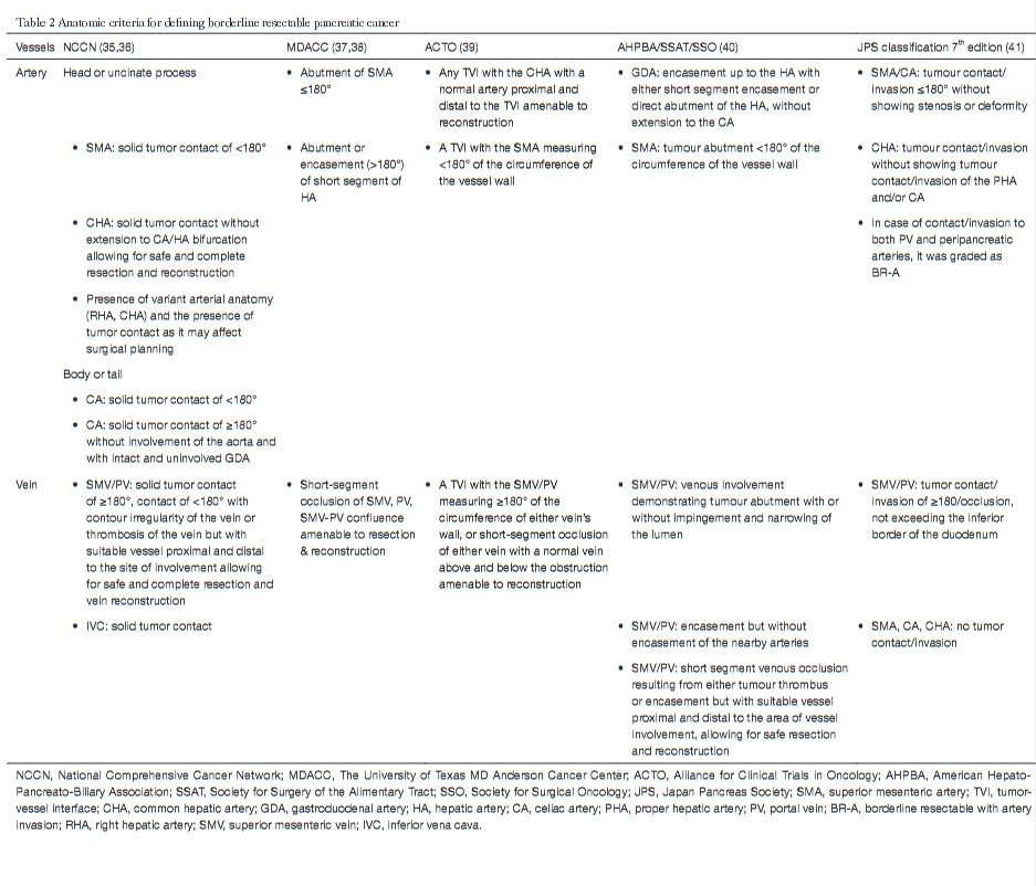
Pancreatic cancer basically divides into resectable disease, borderline resectable disease, unresectable locally advanced disease, and metastatic disease. Resectability for localized disease bases on the extent of adjacent arterial and venous involvement by tumor. For resectable pancreatic cancer, radical resection of pancreatic cancer with adjuvant systemic chemotherapy currently remains the standard treatment approach and is the only chance for long-term survival. A borderline resectable pancreatic cancer (5–10%) is a tumor with limited vascular involvement that is technically resectable but with a high chance of positive resection margins. Many groups have proposed various definitions of borderline resectable pancreatic cancers (35–41) (Table 2). The optimal treatment strategy for borderline resectable pancreatic cancer is still uncertain. Traditionally, “upfront surgery” is the main treatment of choice. Identifying borderline resectable disease is important because a vascular resection +/− reconstruction is more likely needed at the time of pancreatoduodenectomy, and instead of a traditional “upfront surgery” approach, these patients may benefit from neoadjuvant treatment to increase the chance of R0 resection and decrease the surgical morbidity. However, the role and advantages of neoadjuvant treatment in resectable pancreatic cancer and borderline resectable pancreatic cancer remains unclear. There is increasing evidence showing benefits of neoadjuvant therapy in borderline resectable pancreatic cancer. For unresectable locally advanced pancreatic cancer, and metastatic disease, systemic chemotherapy is the choice of treatment. Surgical resection can be considered for those down-staged from unresectable locally advanced disease to resectable disease.
Surgery
For resectable pancreatic cancer, radical resection of pancreatic cancer with adjuvant systemic chemotherapy remains the current standard treatment approach and is the only chance for long-term survival, even there is a high rate of recurrence (42). However, only small proportion of patients (10–20%) with pancreatic cancer are found to be resectable during the diagnosis.
Systemic therapy for metastatic pancreatic cancer
At the time of diagnosis, approximately 50% of patients with pancreatic cancer present with distant metastases. Systemic therapy remains the primary treatment modality aiming at palliating symptoms and prolonging survival.
Conclusions
Pancreatic cancer is a challenging disease for management. Radical surgery itself is not enough for prolong survival. The improvement of chemotherapy, target agents and immunotherapy with multidisciplinary approach for pancreatic cancer patients will be the only solution for improvement of their quality of life and survival outcomes.
The adoption of adjuvant treatment for operable disease and systemic treatment for inoperable disease is based on the published randomized trials. The adoption of neoadjuvant treatment and new treatment modalities such as target agents and immunotherapy is still limited by small number of studies with favorable outcomes. In the coming time, more well-designed large-scale studies are needed to give more evidence for the literature.