
This post contains links to and excerpts from ESC working group position paper on myocardial infarction with non-obstructive coronary arteries [PubMed Abstract] [Full Text HTML] [Full Text PDF]. Eur Heart J. 2017 Jan 14;38(3):143-153. doi: 10.1093/eurheartj/ehw149.
Here are excerpts from the above article:
Definitions
Rationale for diagnosis
As the name implies, the formulation of the MINOCA diagnosis requires both clinical documentation of an AMI and demonstration of non-obstructive coronary arteries (Table 1).
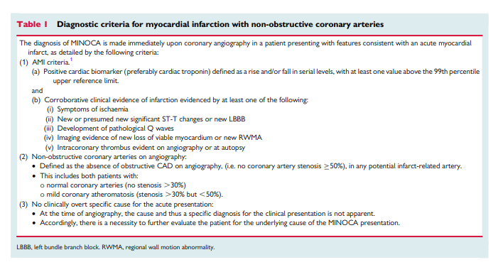
Therefore, if there is no clinically apparent alternative diagnosis, such as myocarditis or pulmonary embolism, the diagnosis of MINOCA is applicable. The rationale for establishing MINOCA as a clinical diagnosis includes: (i) providing a common nomenclature for this group of patients who are often overlooked in contemporary clinical practice, (ii) encouraging routine evaluation for underlying causes in
these patients, and (iii) stimulating further studies into its responsible mechanisms, outcomes, and most appropriate management strategies. Myocardial infarction with non-obstructive coronary arteries should be considered as a ‘working diagnosis’, analogous to heart failure, and thus prompts further evaluation regarding its underlying mechanism(s).
We need to remember that we can have elevated troponins in other clinical scenarios besides acute myocardial infarction.
There are three central concepts in interpreting troponins in the
context of a coronary angiogram showing non-obstructive coronary arteries. First, it should be appreciated that cardiac troponins are ‘organ specific’ and not ‘disease specific’. An elevated cardiac troponin is not necessarily indicative of an AMI but reflects myocardial injury or necrosis. One example of a disease process causing myocardial injury and troponin elevation without an AMI is pulmonary embolism. Thus, there must be corroborative clinical evidence in addition to elevated cardiac troponins to establish the diagnosis of AMI, including MINOCA. However, there is no imaging technology, including cardiac magnetic resonance (CMR) imaging, which can definitively exclude an ischaemic cause of troponin elevation (see ‘MINOCA with normal cardiac MR imaging’). Only a pathologic examination is definitive. Second, rarely the troponin assays may provide spurious results due to analytical issues such as heterophilic antibodies.11 Finally, there are several differential diagnoses for MINOCA, which may arise from both coronary and non-coronary mechanisms as listed in Table 2 and summarized later in this paper. Certainly, the presence of coronary atherosclerotic obstructions, does not exclude other non-cardiac causes of troponin rise.

Angiographic criteria
The angiographic criteria for ‘non-obstructive coronary arteries’ detailed in the MINOCA definition utilizes the conventional cut-off of <50% stenosis, which is consistent with contemporary angiographic guidelines.12 This conventional threshold is somewhat arbitrary and there is substantial inter- and intra-observer variability in visual estimation of angiographic stenosis.
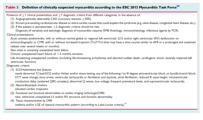
Given the importance of the clinical context in making a diagnosis of MINOCA and the consideration that it is a ‘working diagnosis’, it is not appropriate to use this label when a specific clinical diagnosis is apparent. For example, a young person with a recent viral illness presenting with positional chest pain, diffuse ST elevation, troponin elevation, and found to have normal angiography should be diagnosed as clinically suspected myocarditis with or without associated pericarditis according to ESC 2013 Task Force criteria (Table 3) rather than MINOCA.
Clinical assessment
As stated above, MINOCA is a working diagnosis and should lead the treating physician to investigate underlying causes, analogous to heart failure. This section outlines suggestions for diagnostic testing in order to identify or exclude potential aetiologies discussed in ‘Differential diagnosis’. Left ventriculography or echocardiography should be performed in the acute setting to assess wall motion. This will aid the clinician in determining whether takotsubo cardiomyopathy* is a diagnostic consideration.
*Takotsubo Cardiomyopathy from emedicine.medscape
Cardiac magnetic resonance imaging is the key diagnostic tool tobe employed in MINOCA patients. Late gadolinium enhancement (LGE), when present, permits localization of the area of myocardial damage and provides insight into mechanisms. For example, an area of LGE in the subendocardium suggests an ischaemic cause of injury,
although it does not identify the particular cause of ischaemia (plaque disruption, vasospasm, thromboembolism, or dissection), while a sub-epicardial localization speaks in favour of cardiomyopathy. In other patients, a non-ischaemic appearance of LGE may suggest a diagnosis of myocarditis or an infiltrative disorder. Imaging for myocardial oedema and contractile function may also help localize the area of injury, though with less mechanistic insight.Intracoronary imaging at the time of cardiac catheterization with intravascular ultrasound (IVUS) or optical coherence tomography (OCT) may be useful to identify atherosclerotic plaque disruption and plaque erosion as well as coronary dissection or thrombosis, which may not have been appreciated during angiography. Further research is needed to understand the potential benefit of routine application of intracoronary imaging at the time of coronary catheterization in patients with MINOCA. Coronary CT angiography is another possibility to obtain further information regarding underlying atherosclerosis after the acute angiogram but does not identify plaque rupture or erosion.
We recommend that clinicians consider pulmonary embolism as
a possible cause of myocardial damage and exclude this diagnosisvwith D-dimer testing (usually however also elevated in the setting of AMI) and/or computed tomography (CT) pulmonary angiography as appropriate.17However, no pulmonary embolism was found among 100 consecutive patients with MINOCA who underwent CT pulmonary angiography in one series.18 Furthermore, it is important to consider type-2 AMI1 causes, in which a condition other than coronary plaque instability contributes to an imbalance between myocardial oxygen supply and demand and cause myocardial necrosis, such as tachyarrhythmia, haemorrhage, sepsis, and hypertensive crisis, as potential causes of MINOCA.
After considering clinically apparent diagnoses, the most common causes of MINOCA that the treating clinician must consider are:
- plaque rupture or erosion,
- coronary artery spasm,
- thromboembolism,
- coronary dissection,
- takotsubo cardiomyopathy,
- unrecognized myocarditis,
- and other forms of type-2 myocardial infarction.
Aetiologic differential diagnosis
Plaque disruption
Atherosclerotic plaque disruption is a frequent cause of MINOCA. Plaque disruption is comprised within type-1 AMI in the Universal Definition of Myocardial Infarction, even when no thrombus can be found.1 Within the Universal definition document, MINOCA comprises 5–20% of all type-1 AMI cases Within the Universal definition document, MINOCA
comprises 5–20% of all type-1 AMI cases. The term disruption encompasses imaging and pathologic findings of plaque rupture, ulceration, or erosion. Intraplaque haemorrhage may also play a role. Two independent studies using intravascular ultrasound identified plaque rupture or ulceration in 40% of patients with MINOCA.19,20 . . . . Plaque erosion has also been reported in MINOCA and is characterized by thrombus superimposed on plaque with an intact fibrous
cap or without a fibrous cap.Myonecrosis in MINOCA with plaque disruption is mediated by
thrombosis, thromboembolism, superimposed vasospasm, or a
combination of these processes. One theory that has been proposed as an explanation for MINOCA in the presence of verified myocardial infarction is spontaneous thrombolysis or autolysis of a coronary thrombosis. Spontaneous thrombolysis is thought to be an endogenous protective mechanism against thrombus formation even in the presence of a ruptured coronary plaque.23 Indeed, CMR imaging may show large areas of myocardial oedema with or without small areas of necrosis among patients with MINOCA and plaque disruption, suggesting that flow was compromised transiently in a larger vessel.19 The theory that spontaneous coronary thrombolysis rather than vasospasm leads to this appearance can neither be dismissed nor proved, and both may play a role. In other cases of MINOCA with plaque disruption, CMR imaging shows a
smaller, well-defined area of LGE, subtended by a smaller vessel, suggesting that embolization of atherothrombotic debris from the disruption site is the most likely mechanism of myonecrosis.19Thrombosis and/or thromboembolism almost certainly play a
major role in pathogenesis of MINOCA with plaque disruption.
Therefore, dual antiplatelet therapy is recommended for 1 year followed by lifetime single antiplatelet therapy for patients with suspected or confirmed plaque disruption and MINOCA.3 Because disruption occurs on a background of non-obstructive CAD, statin therapy is also recommended even if only a minor degree of atherosclerosis is found.The prognosis of patients with plaque disruption as the cause of
MINOCA has not been investigated in comparison with other subtypes of MINOCA. However, the finding of plaque rupture on OCT was associated with major adverse cardiac events in a cohort of patients undergoing OCT for acute coronary syndrome.10Coronary artery spasm
Coronary artery spasm may potentially contribute to the pathogenesis of AMI in patients with obstructive CAD and particularly warrants close consideration in those with MINOCA. It reflects a vascular smooth muscle hyper-reactivity to endogenous vasospastic substances (as in vasospastic angina) but may also occur in the context of exogenous vasospastic agents (e.g. cocaine or metamphetamines).30 Provocative spasm testing has demonstrated inducible spasm in 27% of patients with MINOCA suggesting that it is a common and an important pathogenetic mechanism in MINOCA.31 Considering that nitrates and especially calcium channel blockers are effective therapies for coronary artery spasm, with the latter shown to prevent cardiac events in vasospastic angina, this diagnosis and treatment needs to be carefully contemplated.32,33
Myocardial infarction with non-obstructive coronary arteries may be the de novo presentation for patients with vasospastic angina, or an interim event in those with the chronic established form of the disorder. Clinical features of vasospastic angina that may allude to the diagnosis in patients with MINOCA include recurrent episodes of rest angina that promptly respond to short-acting nitrates, especially if associated with transient ischaemic ECG changes and demonstrating a circadian pattern (typically as nocturnal angina). Thus, a diagnosis of vasospastic angina can be made if spontaneous episodes of rest angina are associated with ST-segment changes that respond promptly to short-acting nitrates. However, spontaneous episodes are infrequently documented therefore requiring provocative spasm testing to be undertaken if the diagnosis is to be pursued. . . . Microvascular spasm is also a potential cause of MINOCA since elevated troponins have been detected via ultrasensitive assays following provocative spasm testing, despite the absence of inducible large vessel spasm.35
Coronary thromboembolism
Thrombosis may be a contributory mechanism to AMI in the setting of plaque disruption or coronary artery spasm, or may be the cause of MI in the absence of these factors. Coronary thrombosis may arise from hereditary or acquired thrombotic disorders and coronary emboli may occur from coronary or systemic arterial thrombi. Hereditary thrombophilia disorders include Factor V Leiden thrombophilia, Protein S and C deficiencies. Thrombophilia screening studies in patients with MINOCA have reported a 14% prevalence of these inherited disorders.31 Acquired thrombophilia disorders should also be considered such as the antiphospholipid syndrome and myeloproliferative disorders, , although these have not been systematically investigated in MINOCA.
Coronary emboli may occur in the context of the above thrombophilia disorders or other predisposing hypercoagulable states such as atrial fibrillation and valvular heart disease. Emboli may arise from non-thrombotic sources also including valvular vegetations, cardiac tumours (e.g. myxoma and papillary fibroelastoma), calcified valves, and iatrogenic air emboli.
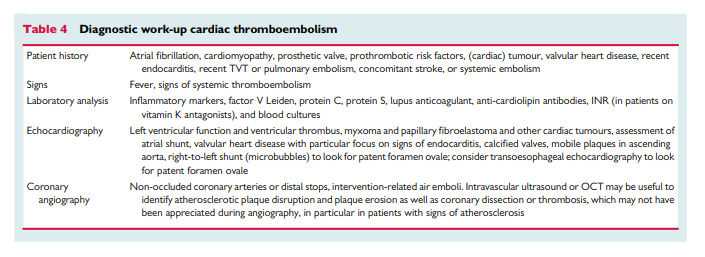
The clinical assessment of coronary thromboembolic disorders,
including paradoxical embolism, as a potential cause of MINOCA is summarized in Table 4. Although the prevalence of coronary thromboembolism in MINOCA is thought to be low, this in part may be due to inadequate screening. For example, pruned small coronary vessels obstructed with thrombi or emboli may be missed on angiography. Aortic valve disease (ectatic calcification, vegetation, or papillary fibroelastoma) may not be noted and thrombophilia disorders may not even be assessed. The importance of identifying these potential causes is the availability of targeted therapies, although their use in the context of MINOCA is limited.
Coronary dissection
Spontaneous coronary dissection typically causes an AMI via luminal obstruction, although this may not always be apparent on coronary angiography, prompting a diagnosis of MINOCA.38 Intramural haematoma of the coronary arteries without intimal tear presents similarly.39 Intracoronary imaging is pivotal in making this diagnosis.40 The condition is more common among women. The reasons for the occurrence of coronary dissection are still unclear but fibromuscular dysplasia* is present in other vascular beds in the majority of cases when screening is performed.41 Changes in the intima-media composition due to hormones, pregnancy, and delivery have also been implicated. Most dissections occur in the absence of atherosclerotic disease and in these cases, statin therapy is not recommended.42 A conservative management approach is advocated because coronary intervention and stenting tend to cause propagation of the dissection and outcomes are acceptable with medical management.43
*See Fibromuscular Dysplasia in emedicine.medscape.com
Coronary dissection
Spontaneous coronary dissection typically causes an AMI via luminal obstruction, although this may not always be apparent on coronary angiography, prompting a diagnosis of MINOCA.38 Intramural haematoma of the coronary arteries without intimal tear presents similarly.39 Intracoronary imaging is pivotal in making this diagnosis.40 The condition is more common among women. [And the etiology is unclear.]
Takotsubo cardiomyopathy
Takotsubo cardiomyopathy44 often presents as an acute coronary syndrome with ST segment changes.45,46 The transient nature of left ventricular dysfunction has puzzled physicians worldwide.47 Clinical presentation is characterized by acute, reversible heart failure associated with myocardial stunning, in the absence of occlusive CAD.48 The prognosis is generally good although several studies have demonstrated significant complications in the acute phase49 – 52 and more studies with long-term follow-up are required.
Distinguishing takotsubo cardiomyopathy from acute myocarditis and AMI due to occlusive CAD may be challenging.
Cardiac magnetic resonance imaging performed early after admission to hospital will in many cases help to establish the diagnosis based on a typical pattern of oedema, emphasizing the importance of performing CMR in MINOCA.
Myocarditis
Since clinical presentation is polymorphic, the 2013 ESC Task Force has introduced rigorous criteria for clinically suspected myocarditis36 that are outlined in Table 3 [Above]. Certain diagnosis of myocarditis and of its specific aetiopathogenetic forms can only be achieved by endomyocardial biopsy (EMB).36
The prevalence of myocarditis among patients with a clinical diagnosis of MINOCA varies based on the populations studied, with a prevalence of 33% in a recent meta-analysis.55 The most common cause of biopsy-proven myocarditis is viral infection, confirmed with polymerase chain reaction (PCR) assay of the pathogen DNA/RNA on EMB. Other causes of myocarditis are immunemediated diseases, endocrine diseases, drugs, and toxins.36,56 – 58
Autoimmune myocarditis may occur with exclusive cardiac involvement or in the context of systemic autoimmune disorders, e.g. systemic lupus erythematosus and is infection-negative by PCR on EMB.36,59,60The initial investigation of suspected myocarditis should include
CMR imaging. Although this non-invasive investigation compares favourably with the gold-standard technique of EMB,36,56,61,62 only EMB provides the opportunity of identifying the underlying cause for the myocarditis. Lurz et al.63 reported that CMR imaging detected 79% of EMB-confirmed myocarditis. Also, in the new ESC guidelines on Pericardial disease CMR is recommended for the confirmation of myocardial involvement (myocarditis) as a Class I recommendation.64The importance of diagnosing myocarditis in patients with MINOCA relates to its prognosis and treatment. Although myocarditis resolves over a 2 –4 weeks period in 50% of patients, 12–25% may acutely deteriorate and either sucumb to fulminant heart failure or progress onto end-stage dilated cardiomyopathy requiring heart transplantation.36
Other forms of type-2 acute myocardial infarction
Type 2 AMI is defined as myocardial cell necrosis due to supply –demand mismatch, characterized by significant increase and/or decrease in troponins with at least one value above the 99th percentile of a normal reference population in the absence of evidence for coronary plaque rupture in addition to at least one of the other criteria for AMI.1 Major determinants of myocardial oxygen demand include systolic wall tension, contractility, and heart rate, while myocardial oxygen supply is conveyed by coronary blood flow and oxygen content. Conditions underlying type-2 AMI include anaemia, tachy- brady-arrhythmia, respiratory failure, hypotension, shock, severe hypertension with or without left ventricular hypertrophy, severe aortic valve disease, heart failure, cardiomyopathy, and injurious effects of toxins (e.g. sepsis) and pharmacological agents (e.g. catecholamines).65 Importantly, all these conditions may also unmask underlying obstructive CAD. Among patients with non-obstructive CAD, a profound supply – demand mismatch should be present to consider type-2 AMI. Therapeutically, the condition underlying the oxygen supply –demand mismatch is to be reversed if possible.
Myocardial infarction with non-obstructive coronary arteries of uncertain aetiology
Cardiac magnetic resonance imaging criteria
Cardiac magnetic resonance imaging is a useful tool in MINOCA patients because it not only provides insights into potential causes but may also provide confirmation of the diagnosis of AMI. In particular, the presence and pattern of any LGE may point towards a vascular or non-vascular cause. However, 8–67% of patients with MINOCA have no evidence of LGE, myocardial oedema, or wall motion abnormalities on CMR.18,19,68 – 74 The MINOCA subgroup with normal CMR raises the question whether the raised troponin was a marker of myocardial injury or perhaps related to an alternate diagnosis.
When CMR is normal and diagnostic evaluation as recommended herein does not reveal the mechanism of AMI, there is a diagnostic and therapeutic dilemma for clinicians. Unfortunately, there are no systematic investigations addressing this issue.
[IVUS (for plaque rupture and dissection) and testing for spasm and microvascular disease are not routinely performed. Thus some etiologies can be undiagnosed presenting a diagnostic dilemma.]
An alternative consideration is that the troponin rise is not indicative of AMI and is instead due to other causes such as pulmonary embolism or myocarditis. These alternate causes should be reconsidered when CMR is normal.
In the absence of systematic evaluation of underlying
mechanisms and clinical characteristics, any treatment recommendations remain empiric. We propose aspirin, statins and, in
cases of vasospasm, calcium channel blockers as routine treatments since these would be of benefit for the potential underlying mechanisms of coronary plaque disruption, coronary spasm, and thromboembolism.Knowledge gaps
We have highlighted that MINOCA is a heterogeneous entity with many potential aetiologies that need to be elucidated by an accurate and stringent commonly agreed diagnostic algorithm (Figure 1).

Summary
Myocardial infarction with non-obstructive coronary arteries is a heterogeneous entity with a prevalence of 1 – 13% of all patients with a clinical diagnosis of AMI. There are several potential aetiologies that should be elucidated by a commonly agreed diagnostic algorithm, proposed herein. Rational treatment follows from an aetiologic diagnosis, since therapy that may be appropriate for one cause (e.g. anticoagulation for thromboembolism or calcium channel blockers for vasospasm) will not be appropriate for all MINOCA patients. In MINOCA patients without an obvious aetiology after initial evaluation including echocardiography, we recommend a routine examination with CMR imaging. Multi-centre clinical trials
of diagnostic and therapeutic strategies are needed. These results will have great impact on both treatment and prognosis of these patients.


